

CD Molecule Nomenclature: New CD Markers and HLDA Updates
Table of Contents
- How CD Nomenclature Works
- HLDA and HCDM
- How a New CD Number Is Assigned
- CD vs CDw Designations
- Latest CD Molecules From HLDA11
- New GPCR CD Molecules
- Updated CD Marker Table
- Why New CD Designations Matter
- Applications in Flow Cytometry and Antibody Development
- Available Recombinant CD Molecules
- Frequently Asked Questions
Cluster of differentiation (CD) nomenclature provides a standardized system for identifying cell-surface molecules involved in immune cell development, differentiation, activation, and communication. Originally introduced to organize monoclonal antibodies according to their recognition of leukocyte antigens, the CD system has evolved into an internationally recognized framework for naming molecular targets in immunology, hematology, cancer biology, and antibody research.
Well-established CD markers such as CD3, CD4, CD8, CD19, and CD45 remain essential for immune cell identification. However, advances in molecular profiling, antibody validation, and high-dimensional immunophenotyping continue to expand the CD molecules list beyond conventional lineage markers. Newly characterized receptors, including certain G protein-coupled receptors (GPCRs), are increasingly incorporated into the standardized CD nomenclature system.
The Human Leukocyte Differentiation Antigens (HLDA) Workshops and the Human Cell Differentiation Molecules (HCDM) organization play central roles in evaluating candidate antigens, validating antibodies, and assigning new CD designations. Recent HLDA11 developments have highlighted the importance of GPCRs in immune cell trafficking, tissue localization, and inflammatory signaling.
This article explains how CD nomenclature works, how new CD numbers are assigned, what distinguishes CD from CDw designations, and which GPCR molecules received new nomenclature in HLDA11. It also examines the implications for flow cytometry, antibody development, and recombinant CD antigen research.
Explore Our CD Antigen Products
Learn More
How CD Nomenclature Works
CD nomenclature was developed to address a major challenge in immunological research: different laboratories frequently generated monoclonal antibodies against the same leukocyte surface molecules but used inconsistent names for these reagents.
Without a standardized system, comparing antibody specificity, immune cell expression patterns, and experimental findings across research groups was difficult.
The CD system introduced a systematic approach to grouping antibodies based on their antigen recognition and subsequently identifying the molecular targets associated with each cluster.
From Monoclonal Antibody Clusters to Molecular Identity
Early CD designations were based primarily on antibody recognition patterns and experimental comparisons of leukocyte antigens. As protein sequencing and molecular cloning techniques advanced, these designations increasingly became associated with defined genes and proteins.
Today, a CD number generally refers to a characterized molecule or molecular complex rather than a specific monoclonal antibody clone.
For example, CD3 identifies a T-cell receptor-associated signaling complex, while CD19 refers to a B-cell surface glycoprotein involved in signaling regulation. CD45, encoded by PTPRC, is a receptor-type protein tyrosine phosphatase expressed on most nucleated hematopoietic cells.
However, the relationship between a CD designation and a gene is not always one-to-one. Some CD numbers encompass related protein isoforms, molecular complexes, or receptor variants.
Table 1. Representative CD Molecules and Their Biological Roles
| CD designation | Molecular identity | Major biological function |
|---|---|---|
| CD3 | T-cell receptor-associated CD3 complex | TCR signal transduction |
| CD4 | CD4 glycoprotein | T-cell co-receptor activity |
| CD8 | CD8α/CD8β co-receptor system | Cytotoxic T-cell antigen recognition |
| CD19 | CD19 glycoprotein | B-cell signaling and identification |
| CD20 | MS4A1 | B-cell development and signaling regulation |
| CD25 | IL2RA | High-affinity IL-2 receptor component |
| CD45 | PTPRC | Leukocyte signaling regulation |
| CD69 | CD69 | Early lymphocyte activation and tissue retention |
| CD80 | CD80 | T-cell costimulation |
| CD86 | CD86 | Antigen-presenting cell costimulation |
| CD274 | CD274 / PD-L1 | Immune checkpoint regulation |
These markers are commonly used to establish immune cell identities and investigate functional changes associated with activation, differentiation, and disease.
Researchers can explore additional CD antigens and immune cell surface markers to understand how different receptor families contribute to immune cell phenotyping and functional studies.
CD Numbers Are Not Functional Classifications
CD numbers do not necessarily indicate molecular function, evolutionary relationships, or the cellular lineage in which a molecule is expressed.
For example, CD4 and CD8 are both T-cell co-receptors, but their numerical proximity does not reflect a shared molecular structure. Similarly, proteins with widely separated CD numbers may belong to closely related functional pathways.
A CD molecule may also be expressed by several immune cell populations rather than a single lineage.
CD4 is strongly associated with helper T cells but can also be expressed by certain myeloid populations. CD16 is found on natural killer cells and subsets of monocytes, neutrophils, and other immune cells.
Consequently, accurate immunophenotyping generally requires combinations of markers rather than reliance on a single CD designation.
The expanding cluster of differentiation molecules list provides a useful framework for selecting complementary markers in immune profiling studies.
HLDA and HCDM
The Human Leukocyte Differentiation Antigens Workshops were established to coordinate the characterization of leukocyte surface antigens and the monoclonal antibodies that recognize them.
The first HLDA Workshop was held in Paris in 1982. Its central objective was to compare antibodies developed by independent laboratories and determine whether they recognized the same or different leukocyte molecules.
The workshop approach introduced a collaborative system in which antibody reagents could be evaluated under standardized conditions across participating research groups.
Over subsequent decades, HLDA workshops helped establish internationally recognized CD designations and improve the consistency of immune cell marker identification. [1]
What Is the Role of HLDA?
HLDA workshops evaluate candidate molecules and their corresponding antibodies using molecular and cellular evidence.
Major activities include:
- Characterizing candidate cell-surface antigens and their molecular identities.
- Assessing monoclonal antibody recognition and specificity.
- Comparing antigen expression across immune cell populations.
- Evaluating reproducibility between independently developed antibody reagents.
- Supporting the assignment and refinement of CD nomenclature.
While early workshops concentrated primarily on traditional leukocyte differentiation antigens, more recent initiatives have incorporated receptors and surface proteins with broader immunological functions.
These include signaling receptors, adhesion molecules, costimulatory proteins, and GPCRs involved in immune cell migration and inflammatory responses.
What Is HCDM?
Human Cell Differentiation Molecules (HCDM) is the international organization associated with the HLDA workshop program and the continued development of standardized CD nomenclature.
HCDM supports scientific collaboration surrounding the characterization of cell-surface molecules, the evaluation of antibody reagents, and the assignment of CD designations.
The organization contributes to maintaining a standardized nomenclature framework that can accommodate advances in molecular immunology and cell-surface receptor research.
Importantly, not every newly discovered membrane protein automatically receives a CD number. The assignment process considers the molecular identity of the target, the available validation evidence, and its suitability for inclusion in the CD nomenclature framework.
How a New CD Number Is Assigned
The identification of a previously uncharacterized immune cell-surface receptor does not automatically result in a new CD designation.
Before a molecule can be incorporated into the nomenclature system, its molecular identity and the specificity of relevant antibody reagents must be sufficiently characterized.
Although individual workshop procedures can vary, the evaluation process can be summarized in five major stages.
1. Candidate Antigen Identification
Potential new CD molecules may be identified through transcriptomics, proteomics, genome-wide analyses, receptor screening, and monoclonal antibody development.
For example, single-cell RNA sequencing can reveal receptors preferentially expressed by particular immune cell populations.
However, transcript detection alone is insufficient to confirm cell-surface protein expression. Receptor abundance can be affected by translation efficiency, intracellular trafficking, protein turnover, and ligand-induced internalization.
Additional protein-level evidence is therefore important.
2. Monoclonal Antibody Generation and Screening
Candidate antibodies are evaluated for their ability to recognize the target molecule under relevant experimental conditions.
Antibodies that detect the native receptor on intact living cells can be particularly valuable for flow cytometry and cell-surface immunophenotyping.
For certain molecular families, including GPCRs, antibody recognition may depend on the receptor's native three-dimensional conformation.
The use of appropriately designed recombinant CD antigen proteins can support antibody screening and target-binding characterization, although cell-based validation is often necessary.
3. Molecular Identity and Specificity Validation
The relationship between antibody recognition and the intended molecular target must be established.
Useful methods may include gene knockout, gene overexpression, immunoprecipitation, receptor-binding assays, and comparison with independently characterized antibodies.
Where related receptors share substantial sequence similarity, cross-reactivity testing is particularly important.
Antibodies that recognize a conserved region within a receptor family may produce apparent positive signals that do not uniquely identify the intended target.
4. Cellular Expression Profiling
Candidate antigens are evaluated across relevant immune cell populations to determine their distribution and expression characteristics.
Flow cytometry, antibody-binding quantification, and genetically defined cell models can provide complementary evidence.
Expression profiling may also reveal differences related to cellular differentiation, activation state, tissue origin, or disease context.
Reproducibility across independent experiments and laboratories strengthens confidence in the biological interpretation of these patterns.
5. Scientific Review and Nomenclature Assignment
The resulting molecular and antibody-validation evidence is evaluated within the relevant HLDA/HCDM nomenclature process.
A molecule may receive an established CD designation, a provisional CDw designation, or remain without a designation pending further evidence.
The following illustration summarizes this conceptual process.
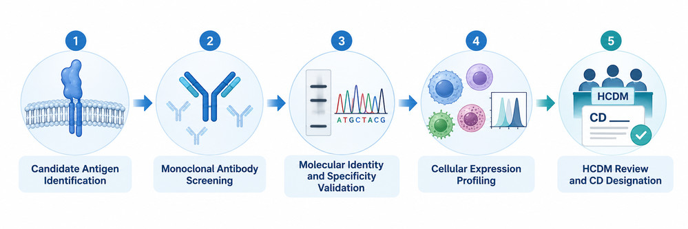 Fig 1. CD Molecule Nomenclature Assignment Workflow
CD vs CDw Designations
An important aspect of CD nomenclature is the distinction between established CD designations and provisional CDw designations.
The lowercase letter w historically refers to the workshop designation system and indicates that the CD assignment is provisional.
A CDw number provides a standardized reference for a molecule while preserving the distinction between provisional and established nomenclature.
Table 2. Differences Between CD and CDw Designations
| Feature | CD designation | CDw designation |
|---|---|---|
| Status | Established | Provisional |
| Prefix | CD | CDw |
| Example | CD375 (GPR15) | CDw376 (GPR26) |
| Molecular identification | Characterized molecular target | Characterized or proposed target with provisional nomenclature |
| Antibody evidence | Evaluated within the nomenclature process | May require additional confirmation |
| Future nomenclature | Generally retained | May be revised or confirmed |
| Recommended reporting | CD number and gene symbol | CDw number and gene symbol |
A provisional designation does not necessarily indicate that the receptor itself is biologically uncharacterized.
For example, GPR183 is a well-studied oxysterol-responsive GPCR involved in immune cell positioning, yet the HLDA11 nomenclature publication identifies it as CDw381.
Similarly, FPR2 participates in inflammatory and inflammation-resolution pathways but carries the provisional designation CDw379 in the same publication.
Researchers should preserve the lowercase w when referring to these molecules until an official nomenclature revision establishes a different designation.
This distinction is particularly relevant when maintaining databases, preparing flow cytometry panels, or documenting antibodies against new CD markers and cell-surface antigens.
Latest CD Molecules From HLDA11
The Eleventh Human Leukocyte Differentiation Antigens Workshop (HLDA11) represents an important development in the characterization of immune cell-surface receptors.
One of its major documented outcomes is the assignment of CD nomenclature to 13 GPCR molecules expressed by immune cells.
The results were reported by Fernández-Calles and colleagues in a 2025 publication in European Journal of Immunology. [2]
The designations include CD198, CD199, CD372–CD375, CD377, CD378, and CD380, together with the provisional CDw376, CDw379, CDw381, and CDw382 identifiers.
These GPCR molecules participate in a variety of biological pathways, including chemokine-mediated migration, lipid mediator signaling, complement activation, and immune cell positioning.
Why GPCRs Were a Focus of HLDA11
GPCRs represent one of the largest families of cell-surface signaling proteins and are important regulators of both innate and adaptive immunity.
Despite their biological significance, many GPCRs have historically been underrepresented in CD nomenclature.
Several factors contribute to this gap.
GPCRs contain seven transmembrane helices and often have relatively small extracellular regions, complicating antibody generation against native conformational epitopes.
Many receptors also display low or variable surface expression on primary leukocytes.
Furthermore, receptor internalization and ligand-dependent regulation can influence antibody recognition and flow cytometry measurements.
HLDA11 addressed these challenges by emphasizing antibody validation and quantitative analysis of GPCR expression across leukocyte subsets. [2]
Explore Our CD Antigen Products
Learn More
Major Outcomes of the HLDA11 GPCR Study
The 2025 publication reported 13 GPCR targets with new CD nomenclature assignments, including nine established CD designations and four provisional CDw designations.
In addition to assigning nomenclature, the study examined antibody specificity and receptor expression patterns across immune cell populations.
These findings can support subsequent investigations into GPCR-mediated immune cell migration, inflammatory signaling, and potential therapeutic target characterization.
However, the HLDA11 GPCR report should not be interpreted as a complete update of every CD molecule in the broader nomenclature system.
Its principal contribution was the characterization and standardized naming of the GPCR targets examined in that workshop study.
New GPCR CD Molecules
The newly designated GPCR molecules include chemokine receptors, complement receptors, lipid mediator receptors, and other signaling proteins.
Each receptor has distinct ligand interactions, expression characteristics, and potential relevance to immune system biology.
CD372 (CCR10): Tissue-Directed Immune Cell Migration
CCR10 is a chemokine receptor that responds to ligands including CCL27 and CCL28.
It contributes to immune cell trafficking in epithelial and mucosal environments and has been investigated in the context of tissue-specific immune responses.
The CD372 designation provides a standardized identifier for linking CCR10 molecular biology with cell-surface immunophenotyping.
CD373 (CX3CR1): Monocyte and Cytotoxic Cell Biology
CX3CR1 is the receptor for CX3CL1, also known as fractalkine.
CX3CR1 is involved in leukocyte trafficking and is relevant to the biology of monocytes, cytotoxic lymphocytes, and tissue-associated immune populations.
Its expression can vary among cellular subsets.
For example, monocyte studies frequently examine CX3CR1 expression alongside CD14 and CD16 to investigate differentiation and trafficking-associated phenotypes.
The CD373 designation helps integrate CX3CR1 into standardized CD marker documentation.
CD374 (XCR1): Dendritic Cell Identification
XCR1 is a chemokine receptor associated with conventional type 1 dendritic cells (cDC1).
It recognizes XCL1 and is relevant to dendritic cell communication with cytotoxic lymphocytes.
XCR1 has attracted particular interest in studies of antigen cross-presentation and dendritic cell-mediated immune responses.
In immunophenotyping experiments, CD374 can contribute to cDC1 characterization when combined with appropriate lineage and dendritic cell markers.
CD375 (GPR15): Mucosal Immune Regulation
GPR15 is a GPCR implicated in immune cell positioning within mucosal tissues.
It has been studied in relation to intestinal immune homeostasis and inflammatory conditions.
Its expression and biological functions may vary according to species, tissue, and immune cell population.
The assignment of CD375 provides a standardized molecular reference for investigations of GPR15-associated immune pathways.
CD378 (C3AR1): Complement-Mediated Signaling
C3AR1 encodes the receptor for complement component C3a.
The C3a–C3AR1 signaling pathway participates in inflammatory responses and can influence immune cell activation and recruitment.
CD378 is particularly relevant to studies investigating complement activation and its interactions with innate immune signaling.
CDw379 (FPR2): Inflammatory and Pro-Resolving Responses
FPR2 belongs to the formyl peptide receptor family.
It recognizes a diverse range of ligands and participates in inflammatory signaling and pathways associated with inflammation resolution.
Its effects are strongly influenced by ligand identity, cell type, and biological context.
CDw379 is a provisional nomenclature designation and should be reported accordingly.
CDw381 (GPR183): Immune Cell Positioning
GPR183, also called EBI2, is an oxysterol-responsive GPCR.
It regulates immune cell positioning within lymphoid tissues and is particularly relevant to B-cell migration and spatial organization.
The CDw381 designation enables researchers to connect GPR183 and EBI2 terminology with the CD marker framework.
CDw382 (F2RL1): Protease-Activated Signaling
F2RL1 encodes protease-activated receptor 2 (PAR2).
The receptor participates in protease-responsive signaling and is involved in inflammatory and tissue-associated cellular responses.
The provisional CDw382 designation introduces a standardized immunological reference for this receptor.
Together, these molecules illustrate the expanding diversity of CD antigens beyond traditional immune lineage markers.
Researchers examining receptor function and antibody interactions can use the broader CD molecule and recombinant CD antigen collection to identify related research targets and investigate their molecular characteristics.
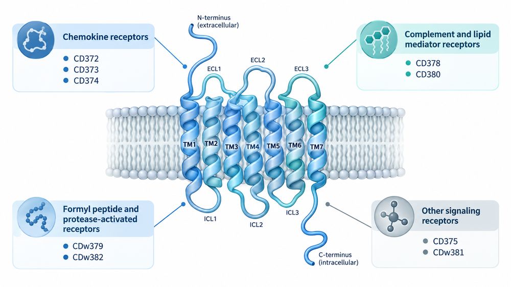 Fig 2. Structure and Classification of Newly Designated GPCR CD Molecules
Updated CD Marker Table
The HLDA11 GPCR nomenclature publication provides an updated reference list connecting CD numbers with their corresponding gene symbols.
This information is useful when comparing experimental studies that use different molecular naming conventions.
Table 3. Updated HLDA11 GPCR CD Molecules List
| CD designation | Gene symbol | Receptor family | Representative research relevance | Nomenclature status |
|---|---|---|---|---|
| CD198 | CCR8 | CC chemokine receptor | Regulatory T cells and tumor immunology | Established |
| CD199 | CCR9 | CC chemokine receptor | Intestinal lymphocyte trafficking | Established |
| CD372 | CCR10 | CC chemokine receptor | Skin and mucosal immune migration | Established |
| CD373 | CX3CR1 | CX3C chemokine receptor | Monocyte biology and immune surveillance | Established |
| CD374 | XCR1 | XC chemokine receptor | cDC1 characterization | Established |
| CD375 | GPR15 | GPCR | Mucosal immune homeostasis | Established |
| CDw376 | GPR26 | Orphan GPCR | Exploratory receptor biology | Provisional |
| CD377 | SSTR3 | Somatostatin receptor | Neuroimmune and signaling research | Established |
| CD378 | C3AR1 | Complement receptor | Complement-mediated inflammation | Established |
| CDw379 | FPR2 | Formyl peptide receptor | Inflammatory and pro-resolving signaling | Provisional |
| CD380 | LTB4R | Leukotriene receptor | Leukocyte migration and lipid signaling | Established |
| CDw381 | GPR183 | Oxysterol-responsive GPCR | B-cell positioning | Provisional |
| CDw382 | F2RL1 | Protease-activated receptor | Protease-dependent inflammatory signaling | Provisional |
Source: Adapted from the nomenclature assignments reported by Fernández-Calles et al., 2025. [2]
The table represents the 13 GPCR molecules documented in the referenced HLDA11 publication. It does not represent the complete CD nomenclature database.
How to Interpret Updated CD Marker Lists
When reviewing updated CD nomenclature, researchers should distinguish three related concepts.
CD Designation
The standardized numerical identifier assigned within the CD nomenclature framework.
Gene Symbol
The standardized genetic identifier corresponding to the molecular target.
Protein or Receptor Name
The descriptive molecular name commonly used in functional or biochemical research.
These identifiers may differ substantially.
For example, CDw381, GPR183, and EBI2 refer to the same molecular target in different naming systems.
Similarly, CDw382 and PAR2 refer to the receptor encoded by F2RL1.
Accurate mapping is especially important when integrating antibody data, flow cytometry results, and transcriptomic datasets.
A comprehensive CD antigens and CD marker reference collection can support research into established surface molecules and their experimental applications.
Explore Our CD Antigen Products
Learn More
Why New CD Designations Matter
The assignment of new CD numbers has implications beyond standardizing molecular names.
New CD designations can improve communication, support reproducibility, and facilitate integration between different immunological research platforms.
Standardized Scientific Communication
A single receptor may be described using a gene symbol, historical protein name, functional name, or laboratory-specific terminology.
This creates challenges when comparing publications and datasets.
A recognized CD designation provides a standardized reference that can be connected to existing molecular identifiers.
For example, documenting CD373 alongside CX3CR1 makes it easier to connect immunophenotyping studies with transcriptomic analyses.
Integration of Transcriptomic and Protein-Level Data
Single-cell RNA sequencing has expanded the identification of cell populations based on gene expression.
However, messenger RNA abundance does not always correspond directly to detectable cell-surface protein levels.
Protein translation, receptor turnover, post-translational modifications, and cellular localization can influence the relationship between RNA and protein expression.
Standardized CD identifiers help connect gene-level information with surface-protein measurements.
This is especially valuable when integrating flow cytometry, mass cytometry, CITE-seq, and related multimodal methods.
Improved Antibody Reagent Documentation
A standardized designation simplifies the annotation of antibodies used to investigate a particular molecular target.
Nevertheless, a CD name alone does not guarantee antibody specificity.
Different antibody clones may recognize distinct epitopes, display different sensitivities, or behave differently under fixation and permeabilization conditions.
Reporting the CD designation together with the target gene, clone identifier, experimental application, and validation information remains important.
Biomarker Discovery and Translational Research
Newly designated CD markers can provide additional options for investigating immune cell states associated with inflammation, tissue migration, and tumor biology.
For example, CCR8 has attracted attention in studies of regulatory T-cell populations in tumor microenvironments.
CX3CR1 is frequently investigated in relation to monocyte differentiation and immune surveillance.
GPR183 contributes to immune cell positioning within lymphoid tissues.
However, CD nomenclature assignment does not independently establish diagnostic or therapeutic utility.
Clinical applications require separate analytical and clinical validation.
Expanded Opportunities for Immune Profiling
New CD designations encourage the evaluation of less commonly studied receptors within established immunophenotyping frameworks.
The integration of conventional lineage markers with newer receptor markers may help researchers identify functional or differentiation-associated differences among cell populations.
For example, pairing CD14 and CD16 with CD373 can provide additional information about monocyte-related phenotypes.
Similarly, CD374 may contribute to dendritic cell characterization when used alongside appropriate lineage and cDC1-associated markers.
Applications in Flow Cytometry and Antibody Development
Flow cytometry remains a major experimental platform for investigating CD molecule expression.
Modern multiparameter and spectral flow cytometry technologies enable researchers to assess increasingly complex combinations of surface markers on individual cells.
The expanding CD nomenclature system provides additional candidate markers for studying immune cell differentiation, migration, and activation.
Flow Cytometry Panel Design
A well-designed immunophenotyping panel generally includes markers that establish the principal identity of the cell population.
Additional markers can then be introduced to investigate differentiation, activation, tissue trafficking, or receptor-specific functions.
For example, CD45 can help identify leukocytes, while CD3, CD19, or myeloid lineage markers distinguish major populations.
Newly designated GPCR CD molecules may then be incorporated to explore more specialized biological features.
Table 4. Example Applications of New CD Markers in Flow Cytometry
| Research application | Core markers | Additional CD markers | Primary objective |
|---|---|---|---|
| Monocyte profiling | CD45, CD14, CD16, HLA-DR | CD373 | Investigate CX3CR1 expression among monocyte subsets |
| Dendritic cell characterization | CD45, HLA-DR, CD11c and lineage exclusion markers | CD374 | Study cDC1-associated phenotypes |
| Regulatory T-cell research | CD3, CD4, CD25, CD127 | CD198 | Examine CCR8-associated Treg populations |
| Mucosal immune research | CD3, CD4 or CD8 | CD199, CD372, CD375 | Investigate tissue-trafficking receptor profiles |
| Innate inflammatory responses | CD45, CD14, CD16 | CD378, CDw379, CD380 | Assess inflammatory receptor expression |
| B-cell positioning research | CD19, CD20, CD27 | CDw381 | Investigate GPR183 expression patterns |
These are illustrative research combinations rather than fully optimized or clinically validated panels. Experimental design, gating strategy, antibody specificity, and appropriate controls must be established for each application.
Antibody Validation for New CD Markers
New CD markers are particularly useful when supported by antibodies that can detect native receptor expression on relevant cell populations.
However, antibody performance may vary depending on receptor structure, antigen presentation, and experimental conditions.
A robust validation strategy may incorporate the following approaches.
Positive and Negative Controls
Positive controls should exhibit characterized target expression, while negative controls help establish nonspecific binding.
Genetically modified cell models, including receptor knockout and overexpression systems, can be especially informative.
Independent Antibody Validation
Where suitable reagents are available, using independent antibody clones can help determine whether observed staining is consistent with target-specific recognition.
Orthogonal Molecular Methods
Flow cytometry findings may be supported by transcript analysis, immunoprecipitation, mass spectrometry, or other appropriate techniques.
Assessment of Related Receptors
Potential cross-reactivity should be evaluated when antibodies are directed against structurally related receptor-family members.
Assay-Specific Controls
Fluorescence-minus-one controls, viability discrimination, compensation or spectral-unmixing controls, and instrument-specific quality controls should be incorporated where appropriate.
Standardized reporting recommendations such as MIFlowCyt support the transparent documentation of flow cytometry experiments. [3]
Explore Our CD Antigen Products
Learn More
Why GPCR Antibody Development Requires Special Consideration
GPCRs are challenging antigens because their functional structures depend on seven transmembrane helices and their interactions with the membrane environment.
Native receptor epitopes may involve multiple extracellular regions that become spatially adjacent only when the protein is correctly folded.
Isolated synthetic peptides or soluble fragments may therefore fail to reproduce the conformational features of the full-length receptor.
Antibodies generated against such fragments can sometimes recognize denatured proteins but show limited binding to native receptors on living cells.
For antibodies intended for flow cytometry or receptor-blocking studies, native-like antigen presentation and cell-based validation may be particularly valuable.
Researchers developing antibodies against recombinant CD molecules and cell-surface antigens should select antigen formats that match the intended biological application.
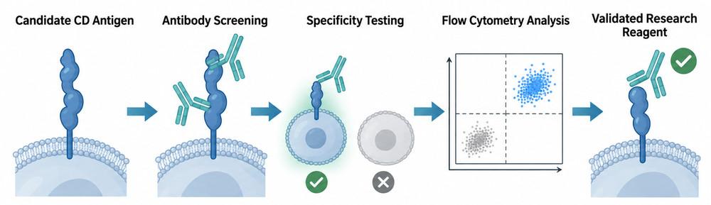 Fig 3. Antibody Validation Workflow for New CD Markers
Available Recombinant CD Molecules
Recombinant CD molecules are useful research materials for studying immune cell-surface receptors, evaluating protein interactions, and developing antibody-based assays.
Depending on the molecular family and experimental objective, recombinant CD antigens may be produced as soluble extracellular domains, tagged proteins, engineered fusion constructs, or full-length membrane-associated proteins.
These different formats offer distinct advantages and limitations.
Applications of Recombinant CD Antigens
Antibody Development and Screening
Recombinant CD antigens can be used to evaluate antibody binding, identify candidate clones, and compare binding specificity.
Purified extracellular domains are commonly used when the protein retains the relevant epitope outside its membrane context.
For conformationally complex targets, particularly GPCRs, full-length or membrane-associated antigen formats may be necessary.
Protein Interaction Studies
Recombinant CD molecules can support biochemical assays investigating receptor–ligand interactions and molecular recognition.
Examples include ELISA, surface plasmon resonance, biolayer interferometry, and other affinity-based analytical techniques.
The reliability of these assays depends on antigen folding, immobilization strategy, and assay conditions.
Immune Receptor Characterization
Recombinant proteins can help characterize molecular interactions involved in immune signaling, receptor recognition, and intercellular communication.
For example, recombinant costimulatory proteins and immune checkpoint molecules can be used in binding studies involving their corresponding interaction partners.
Assay Development and Quality Control
Well-characterized recombinant CD proteins may serve as positive reference materials or assay-development reagents when their identity and performance are appropriate for the experimental system.
Table 5. Recombinant CD Antigen Formats and Applications
| Antigen format | Advantages | Limitations | Typical applications |
|---|---|---|---|
| Soluble extracellular domain | Defined protein sequence and relatively straightforward purification | May not preserve membrane-dependent conformations | ELISA, binding assays, antibody screening |
| His-tagged recombinant protein | Convenient purification and immobilization | Tag position may influence assay behavior | Protein characterization and binding studies |
| Fc-fusion CD protein | Facilitates purification and may enhance apparent binding through avidity | Fc-mediated interactions and dimerization may complicate interpretation | Receptor-binding assays and antibody screening |
| Biotinylated CD protein | Supports controlled immobilization when appropriately designed | Requires suitable labeling strategy | SPR, BLI, capture assays |
| Full-length membrane protein | Includes native receptor sequence and transmembrane regions | Complex extraction and stabilization requirements | GPCR research and membrane receptor studies |
| Receptor-expressing cells | Native-like membrane environment and cellular processing | Background binding and expression variability | Flow cytometry and cell-based antibody screening |
Explore Our CD Antigen Products
Learn More
Key Considerations When Selecting Recombinant CD Proteins
Species and Molecular Identity
Confirm the species, gene symbol, CD designation, and sequence boundaries.
Closely related proteins may display substantial sequence differences that influence antibody recognition.
Expression System
Mammalian expression systems may be appropriate for proteins requiring complex folding and glycosylation.
Bacterial systems can be useful for selected soluble domains, although they may not reproduce native eukaryotic post-translational modifications.
Protein Structure and Functional Activity
The recombinant construct should retain the structural features necessary for the intended assay.
For receptor-binding studies, biological activity or interaction-based quality control may be more informative than protein purity alone.
Fusion Tags and Immobilization
His, Fc, and biotinylation formats may facilitate protein purification, detection, or immobilization.
However, researchers should consider whether the tag influences protein conformation, oligomerization, or apparent binding affinity.
Quality Control
Important quality attributes include identity, purity, molecular integrity, aggregation, and application-relevant functional performance.
Endotoxin levels should also be evaluated when recombinant proteins will be used in endotoxin-sensitive cellular assays.
Recombinant CD Molecules for Immunology Research
Different CD molecule families are useful for different experimental objectives.
T-cell markers such as CD3, CD4, and CD8 are central to lymphocyte research.
Costimulatory molecules such as CD80 and CD86 participate in antigen-presenting cell–T-cell interactions.
Immune checkpoint molecules, including CD274, are important in studies of immune regulation and tumor immunology.
Adhesion molecules and receptors associated with the CD nomenclature system can also support investigations of immune cell trafficking and receptor-mediated signaling.
Researchers can explore the Creative BioMart recombinant CD antigen and cluster of differentiation protein collection to identify relevant molecular targets for antibody development, immune profiling, and protein interaction studies.
Special Considerations for New GPCR CD Antigens
Recently designated GPCR CD molecules may require specialized antigen-production approaches.
Unlike many soluble immunoglobulin-like domains, full-length GPCRs contain multiple hydrophobic transmembrane segments that complicate conventional protein expression and purification.
Preserving a functionally relevant receptor conformation may require detergent-based stabilization, lipid-containing systems, or other membrane-mimetic environments.
Furthermore, an isolated extracellular peptide may be suitable for certain immunization strategies but not for evaluating antibodies that recognize native receptor conformations.
Therefore, researchers should evaluate construct design, native epitope preservation, and antibody application before selecting a recombinant format.
The presence of a new CD designation does not guarantee the availability of a commercial recombinant protein or a validated antibody.
Nevertheless, standardized CD nomenclature helps establish a consistent molecular framework for future antigen development and immunological research.
For related targets, the CD antigen proteins and cell-surface receptor research resources provide a starting point for investigating established CD molecule families and their potential experimental applications.
Frequently Asked Questions
-
What is CD nomenclature in immunology?
CD nomenclature is an internationally recognized system used to identify and classify cell-surface molecules associated with immune cells and other biologically relevant cell populations. It provides standardized identifiers for proteins commonly investigated in immunophenotyping, antibody development, and immune system research.
-
What is the difference between CD markers and CD antigens?
The terms are closely related but emphasize different aspects of the same molecules. CD markers are generally discussed in the context of cell identification and phenotyping, while CD antigens emphasize molecular targets recognized by antibodies. A molecule can function as both a cell-surface marker and an antigen.
-
Who assigns new CD numbers?
New CD designations are assigned through the HLDA/HCDM nomenclature framework. The process draws on molecular characterization, antibody specificity, expression profiling, and scientific review.
-
What new CD molecules were identified in HLDA11?
The 2025 HLDA11 GPCR nomenclature publication reported 13 newly assigned CD names: CD198, CD199, CD372, CD373, CD374, CD375, CDw376, CD377, CD378, CDw379, CD380, CDw381, and CDw382.
-
What does CDw mean?
CDw is a provisional designation in the CD nomenclature system. The lowercase letter w reflects the historical workshop designation convention. It indicates that the assigned CD name is provisional rather than fully established.
-
Why are GPCRs important in new CD marker research?
GPCRs regulate immune cell migration, inflammatory signaling, tissue localization, and responses to extracellular mediators. Their standardized CD designation supports more consistent characterization of receptor expression and antibody specificity.
-
Can new CD markers be used in flow cytometry?
Yes, provided that suitable antibodies have been validated for the intended application. Newly designated GPCR markers may be incorporated into multiparameter flow cytometry panels, but appropriate controls and experimental optimization are required.
-
Are recombinant proteins available for every CD molecule?
No. Recombinant protein availability varies by target, construct type, expression complexity, and supplier. Full-length GPCRs are particularly challenging to produce in native-like forms.
-
How should researchers choose recombinant CD antigens?
Selection should consider species, molecular identity, protein format, expression system, fusion tag, structural integrity, and assay requirements. Antigen choice should match the intended experimental application.
Explore Our CD Antigen Products
Learn More
Conclusion
CD nomenclature remains a fundamental framework for organizing immune cell-surface molecules and improving the consistency of immunological research.
The HLDA11 updates demonstrate how the system continues to evolve as new molecular targets and antibody-validation technologies become available.
The assignment of CD nomenclature to 13 GPCR molecules provides standardized identifiers for receptors involved in chemokine signaling, immune cell migration, complement activation, and inflammatory regulation.
These developments can support more consistent communication across flow cytometry, antibody development, molecular immunology, and receptor biology.
However, accurate interpretation requires careful attention to provisional CDw designations, molecular identity, antibody specificity, and the biological context in which each receptor is studied.
As the list of CD molecules continues to evolve, combining standardized nomenclature with validated antibodies and appropriately characterized recombinant antigens will remain important for reproducible immunological research.
Resource
-
From CD Markers to Immune Maps: A New Era in Immunology
-
The CD Map Is Still Growing: New CD Molecules and HLDA11
-
Antigen Density: Beyond CD-Positive
-
Spatial CD Biology: Why Location Matters in the Tumor Microenvironment
-
Logic-Gated CAR-T: Revolutionizing Cancer Therapy
-
Cluster of Differentiation

References
- Engel P, Boumsell L, Balderas R, et al. CD nomenclature 2015: Human leukocyte differentiation antigen workshops as a driving force in immunology. Journal of Immunology. 2015;195(10):4555–4563. doi: 10.4049/jimmunol.1502033.
- Fernández-Calles J, Kužílková D, Hedin F, et al. CD Molecules Nomenclature 2025: Antibody Validation and Expression Profiling of Immune System G Protein-Coupled Receptors. European Journal of Immunology. 2025;55(12). doi: 10.1002/eji.70099.
- Lee JA, Spidlen J, Boyce K, et al. MIFlowCyt: The minimum information about a flow cytometry experiment. Cytometry Part A. 2008;73A(10):926–930. doi: 10.1002/cyto.a.20623.
- Yáñez-Mó M, Siljander PRM, Andreu Z, et al. Biological properties of extracellular vesicles and their physiological functions. Journal of Extracellular Vesicles. 2015;4:27066. doi: 10.3402/jev.v4.27066.
- Cyster JG, Allen CDC. B cell responses: Cell interaction dynamics and decisions. Cell. 2019;177(3):524–540. doi: 10.1016/j.cell.2019.03.016.

Contact us or send an email at for project quotations and more detailed information.
Quick Links
-

Papers’ PMID to Obtain Coupon
Submit Now -

Refer Friends & New Lab Start-up Promotions