

Myeloid and Dendritic Cell CD Markers for Immune Profiling
Table of Contents
- Why Myeloid Immunophenotyping Requires Marker Panels
- Human Monocyte CD Markers
- Macrophage CD Markers and Phenotypic Plasticity
- Dendritic Cell CD Markers
- Activation, Costimulation and Maturation Markers
- Myeloid CD Markers in the Tumor Microenvironment
- Designing Multiparameter Myeloid Marker Panels
- Common Technical Pitfalls
- Recombinant CD Antigens for Myeloid Research
Why Myeloid Immunophenotyping Requires Marker Panels
Myeloid cells are among the most phenotypically flexible cells of the immune system. Circulating monocytes can enter tissues and differentiate into macrophage-like populations; dendritic-cell subsets share markers with monocytes; and inflammatory signals can substantially alter surface-protein expression.
As a result, there is no single universal "myeloid marker."
Instead, immune profiling uses combinations of CD antigens, HLA molecules, chemokine receptors and exclusion markers.
| Population | Frequently Used Markers | Major Interpretation Issue |
|---|---|---|
| Classical monocytes | CD14 high, CD16 low | Activation can modify phenotype |
| Intermediate monocytes | CD14 high, CD16 positive | Gating boundaries vary |
| Nonclassical monocytes | CD14 low, CD16 high | CD16 also occurs on NK cells and other cells |
| Macrophages | CD68, CD64, CD163, CD206 | Tissue-specific heterogeneity |
| Conventional DCs | CD11c plus subset markers | CD11c is not DC-specific |
| Mature/activated DCs | CD83, CD80, CD86 | Activation context matters |
Human Monocyte CD Markers
Human blood monocytes are commonly separated using CD14 and CD16.
Classical Monocytes
Classical monocytes typically show strong CD14 expression with little or no CD16. They are abundant in peripheral blood and are involved in inflammatory responses, phagocytosis and recruitment to tissues.
Intermediate Monocytes
Intermediate monocytes retain relatively high CD14 while also expressing CD16.
Nonclassical Monocytes
Nonclassical monocytes generally show lower CD14 and higher CD16. They display distinct migratory and vascular-surveillance behaviors.
The CD14/CD16 nomenclature substantially improved standardization, but the boundaries are not biologically absolute. Sample handling, disease state, antibody clone and gating strategy can all affect subset frequencies.
CD64 can provide additional information because it is strongly associated with monocytes and macrophages and represents the high-affinity Fc gamma receptor I.
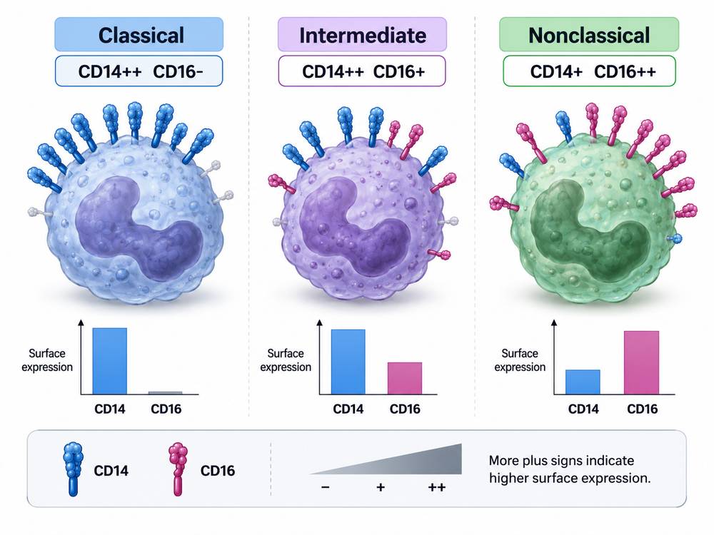 Fig 1. CD14/CD16 Definition of Human Monocyte Subsets
Explore Our CD Antigen Products
Learn More
Macrophage CD Markers and Phenotypic Plasticity
Macrophages are even more heterogeneous than circulating monocytes.
CD68 is one of the best-known macrophage-associated markers. However, much of CD68 is intracellular, and CD68 is not exclusively restricted to macrophages.
CD64 is often useful for identifying differentiated mononuclear phagocytes.
CD163 is a scavenger receptor associated with selected macrophage populations and anti-inflammatory or tissue-repair contexts.
CD206, the mannose receptor, is also frequently used in macrophage studies.
The traditional "M1/M2" classification is useful as an experimental shorthand but does not fully capture macrophage biology in vivo. Tissue macrophages frequently occupy mixed or intermediate transcriptional and functional states.
Consequently, statements such as "CD206-positive means M2 macrophage" are often too simplistic.
A better approach evaluates:
- tissue origin;
- inflammatory context;
- multiple surface markers;
- transcriptional state;
- cytokine production;
- phagocytic function;
- metabolic phenotype.
Dendritic Cell CD Markers
Dendritic cells are specialized antigen-presenting cells that connect innate sensing with adaptive T-cell responses.
CD11c is widely used in DC research, especially in conventional dendritic cells, but it is also expressed by other myeloid populations. Therefore CD11c positivity alone does not establish DC identity.
Modern human DC classification frequently combines HLA-DR with lineage-exclusion markers and subset-associated molecules.
CD83 is particularly important in studies of dendritic-cell activation and maturation. Its expression often increases as DCs mature in response to inflammatory or pathogen-associated signals.
CD80 and CD86 provide additional information about costimulatory capacity.
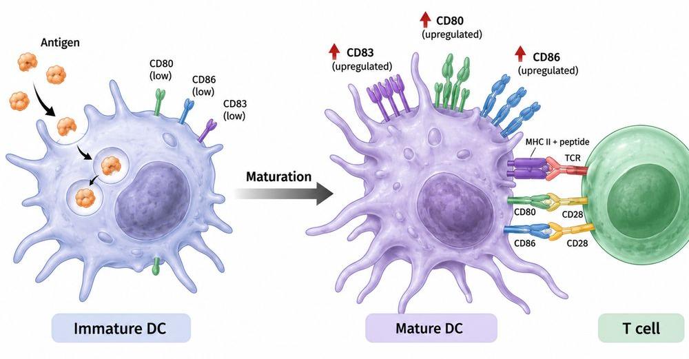 Fig 2. Dendritic Cell Maturation and Costimulation
Activation, Costimulation and Maturation Markers
Myeloid phenotyping becomes especially informative when lineage markers are combined with functional molecules.
| Marker | Primary Use in Myeloid Studies |
|---|---|
| HLA-DR | Antigen-presentation capacity |
| CD80 | Costimulation and APC activation |
| CD86 | Costimulation and APC activation |
| CD83 | Dendritic-cell maturation |
| CD64 | Fc-mediated function, monocyte/macrophage identification |
| CD163 | Macrophage scavenger/reprogramming phenotype |
| CD206 | Tissue macrophage and scavenging-related phenotype |
CD80 and CD86 are particularly important because they bind CD28 and CTLA-4 on T cells and can therefore influence whether antigen presentation leads to effective activation or immune regulation.
This provides a functional bridge between myeloid-cell state and adaptive immunity.
Explore Our CD Antigen Products
Learn More
Myeloid CD Markers in the Tumor Microenvironment
The tumor microenvironment can dramatically remodel myeloid populations.
Tumor-associated macrophages may support or oppose tumor growth depending on context. Myeloid-derived suppressive populations can inhibit lymphocyte activity. Dendritic cells may be numerically present but functionally impaired.
This creates several pitfalls.
A high number of CD68-positive cells, for example, does not automatically indicate a specific functional macrophage state.
Similarly, CD11c-positive cells cannot automatically be classified as functionally competent dendritic cells.
More informative tumor studies combine surface-marker data with spatial localization and functional state.
Modern approaches include:
- high-dimensional flow cytometry;
- spectral flow cytometry;
- mass cytometry;
- single-cell RNA sequencing;
- spatial transcriptomics;
- multiplex immunofluorescence.
CD markers remain especially useful because they permit isolation and experimental manipulation of the cell states discovered by these high-dimensional methods.
Designing Multiparameter Myeloid Marker Panels
A practical panel-development sequence is:
Step 1: Establish sample quality.
Include viability assessment because damaged cells can increase nonspecific binding.
Step 2: Identify leukocytes.
CD45 is commonly used as a broad leukocyte marker.
Step 3: Exclude unwanted lineages.
T-, B- and NK-cell markers can help remove lymphoid populations.
Step 4: Define myeloid lineage.
Use combinations such as CD14, CD16, CD64 and HLA-DR.
Step 5: Separate populations.
Resolve monocyte, macrophage and DC-associated phenotypes.
Step 6: Add functional markers.
CD80, CD83, CD86, CD163 or CD206 can address activation or differentiation questions.
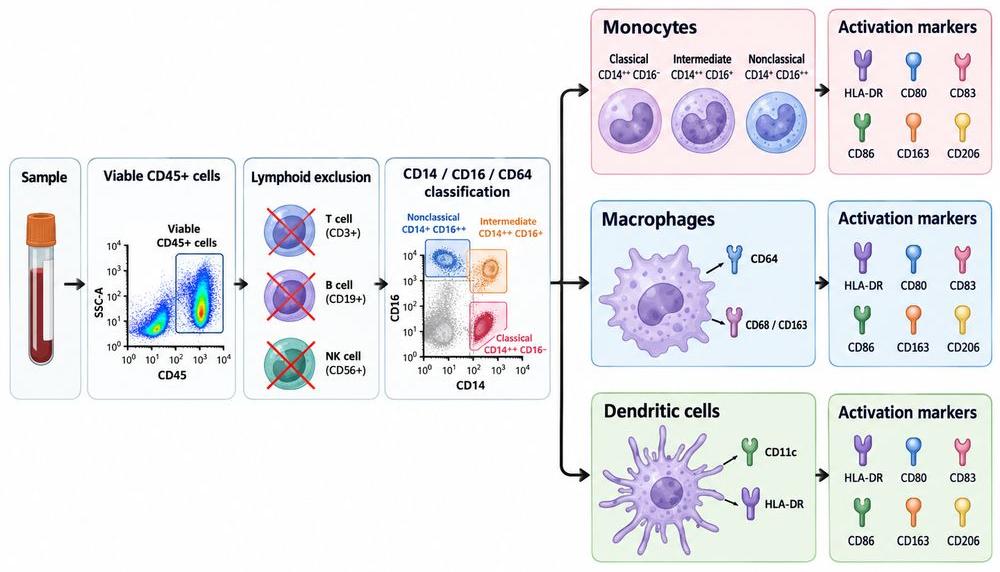 Fig 3. Myeloid Immunophenotyping Workflow
Common Technical Pitfalls
Fc Receptor-Mediated Background
Myeloid cells express multiple Fc receptors. Antibodies can therefore bind through their Fc domains independently of antigen recognition.
Appropriate Fc-blocking reagents and controls are particularly important.
Tissue Digestion
Enzymatic dissociation can alter some surface epitopes. Tissue-processing conditions should therefore be validated.
Marker Modulation
Activation during sample handling can change CD16, CD62L and other surface molecules.
Overinterpretation of One Marker
A marker should be interpreted as evidence contributing to a phenotype, not as an absolute identity.
Explore Our CD Antigen Products
Learn More
Recombinant CD Antigens for Myeloid Research
Well-characterized recombinant CD antigens can support several stages of myeloid-reagent development.
Applications include:
- antibody specificity testing;
- ELISA development;
- receptor-ligand interaction studies;
- antibody competition;
- epitope mapping;
- species cross-reactivity studies.
Targets such as CD14, CD16, CD64, CD80, CD83 and CD86 are particularly relevant to myeloid biology.
When Fc receptor pathways are involved, protein format deserves careful attention. Fc-tagged proteins may interact with Fc receptors independently of the intended binding pair. Appropriate controls or alternative tags may therefore be required.
For translational projects, matched human, mouse and nonhuman-primate CD antigen proteins may support early cross-species evaluation.
The direct answer to the common question "Which CD markers identify myeloid cells?" is therefore:
CD14 and CD16 are commonly used to classify human blood monocytes, while macrophage and dendritic-cell studies typically require broader panels incorporating CD64, CD68, CD163, CD206, CD11c, CD83, CD80, CD86 and HLA-DR. No single CD marker is sufficient for all myeloid populations.
Resource
-
From CD Markers to Immune Maps: A New Era in Immunology
-
The CD Map Is Still Growing: New CD Molecules and HLDA11
-
Antigen Density: Beyond CD-Positive
-
Spatial CD Biology: Why Location Matters in the Tumor Microenvironment
-
Logic-Gated CAR-T: Revolutionizing Cancer Therapy
-
Cluster of Differentiation
References
- Ziegler-Heitbrock L, Ancuta P, Crowe S, et al. Nomenclature of Monocytes and Dendritic Cells in Blood. Blood. 2010;116(16):e74–e80. doi:10.1182/blood-2010-02-258558.
- Li Z, Ju X, Silveira PA, et al. CD83: Activation Marker for Antigen Presenting Cells and Its Therapeutic Potential. Front Immunol. 2019;10:1312. doi:10.3389/fimmu.2019.01312.
- Prechtel AT, Steinkasserer A. CD83: An Update on Functions and Prospects of the Maturation Marker of Dendritic Cells. Arch Dermatol Res. 2007;299(2):59–69. doi:10.1007/s00403-007-0743-z.
- Murray PJ, Allen JE, Biswas SK, et al. Macrophage Activation and Polarization: Nomenclature and Experimental Guidelines. Immunity. 2014;41(1):14–20. doi:10.1016/j.immuni.2014.06.008.

Contact us or send an email at for project quotations and more detailed information.
Quick Links
-

Papers’ PMID to Obtain Coupon
Submit Now -

Refer Friends & New Lab Start-up Promotions