

CD Markers for Immune Cell Immunophenotyping: A Practical Guide
Table of Contents
- What Are CD Markers in Immunophenotyping?
- CD Markers by Immune Cell Type
- Lineage Markers vs Activation Markers vs Differentiation Markers
- Common CD Marker Combinations
- CD Markers in Multicolor and Spectral Flow Cytometry
- CD Antigens as Controls and Standards in Assay Development
- How to Select Recombinant CD Antigens
- Related CD Antigen Products
- Key Takeaways
Immunophenotyping identifies and characterizes immune cell populations according to their expression of cell-surface and intracellular molecules. Among the most widely used immunophenotyping markers are cluster of differentiation, or CD, molecules. By analyzing combinations of CD markers, researchers can distinguish major leukocyte lineages, define developmental and functional subsets, and measure activation, differentiation, exhaustion, or maturation states.
There is rarely a single CD marker that uniquely defines an immune cell population. Instead, reliable CD markers for immune cells are interpreted as combinations. For example, CD3 identifies the T-cell lineage, whereas CD3 together with CD4 or CD8 separates major T-cell subsets. Similarly, CD3 negativity combined with CD56 and CD16 expression is commonly used to characterize NK cells. Multiparameter immunophenotyping studies therefore rely on coordinated marker panels rather than isolated antigens. [1–4]
For assay development, antibody characterization, receptor-binding studies, and other applications, well-characterized CD antigens can also provide defined molecular reagents that complement cell-based controls.
Explore Our CD Antigen Products
Learn More
What Are CD Markers in Immunophenotyping?
The cluster of differentiation nomenclature provides a standardized system for identifying cell-surface molecules recognized by validated monoclonal antibodies. The system emerged from the Human Leukocyte Differentiation Antigen workshops and continues to evolve as additional immune-associated molecules are characterized. The 2025 CD nomenclature update, for example, assigned CD designations to additional immune GPCRs, including CD374 for XCR1. [1]
In flow cytometry, fluorescently labeled antibodies recognize specific CD molecules on individual cells. The resulting expression patterns can be used to answer several different questions:
- What lineage is the cell? Examples include CD3 for T cells and CD19 for B cells.
- Which subset does it belong to? Examples include CD4 versus CD8 T cells and CD14/CD16 monocyte subsets.
- What is its differentiation state? CD45RA, CD45RO, CD27, CD57, and CD197/CCR7 are frequently used for this purpose.
- Is the cell activated? Markers such as CD25, CD38, CD69, HLA-DR, CD137, and CD154 may provide activation-related information.
- How mature or functionally specialized is the cell? Marker combinations can further divide NK cells, dendritic cells, regulatory T cells, memory B cells, and other populations.
Importantly, expression is context dependent. A CD molecule may occur on multiple lineages, and its abundance may change with activation, tissue localization, disease state, or sample processing. Consequently, immune cell surface markers should always be interpreted in the context of the complete phenotype and gating strategy.
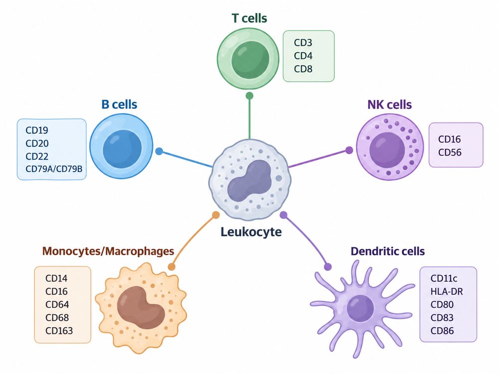 Figure 1. CD Marker Map of Major Immune Cell Types
CD Markers by Immune Cell Type
A practical panel generally begins with lineage-defining markers and then adds subset, activation, and differentiation markers according to the biological question. Broad human PBMC immunophenotyping panels have demonstrated that combinations of CD3, CD4, CD8, CD19, CD56, CD16, CD14, HLA-DR, and related markers can resolve major lymphoid and myeloid populations in a single experiment. [2–4]
| Immune Cell Type | Common Lineage/Identification Markers | Frequently Used Subset or State Markers | Example Phenotype |
|---|---|---|---|
| T cells | CD3 | CD4, CD8, CD25, CD27, CD38, CD45RA/RO, CD69, CD127, CD197 | CD3+CD4+ or CD3+CD8+ |
| B cells | CD19, CD20 | CD21, CD27, CD38, CD24 | CD19+CD20+ |
| NK cells | CD56, CD16 with CD3 exclusion | CD57, CD69, CD94, CD38 | CD3−CD56+ |
| Monocytes | CD14, CD16, HLA-DR | CD64, CD11b | CD14++CD16− or CD14+CD16++ |
| Macrophages | CD64, CD68 | CD163, CD206, HLA-DR | Context- and tissue-dependent |
| Dendritic cells | HLA-DR with lineage exclusion | CD11c, CD1c, CD123, CD141, CD303, CD374/XCR1 | Lin−HLA-DR+ |
These marker profiles represent commonly used starting points rather than universal definitions.
T Cells
CD3 is the primary lineage marker for conventional T-cell immunophenotyping. Once CD3+ cells have been identified, CD4 and CD8 commonly separate helper and cytotoxic T-cell populations.
Additional markers can then resolve functionally important subsets:
- CD4: commonly used to identify helper T cells.
- CD8: commonly used to identify cytotoxic T-cell populations.
- CD25 and CD127: CD4+CD25^high^CD127^low/−^ is a commonly used surface phenotype for enriching regulatory T cells, often supplemented by intracellular FOXP3.
- CD45RA/CD45RO and CD197 (CCR7): widely used to distinguish naïve and memory differentiation states.
- CD27 and CD28: useful in assessing differentiation and co-stimulatory capacity.
- CD38, CD69 and HLA-DR: frequently incorporated into activation panels.
- CD57: associated with advanced differentiation in selected T-cell populations.
One well-established pan-leukocyte strategy evaluates CCR7, CD45RA, CD57, and CD127 for differentiation while assessing CD38 and HLA-DR for activation. [2]
Defined immune cell CD antigens can be useful when validating antibodies or developing binding assays directed against selected T-cell surface proteins.
B Cells
CD19 is one of the most frequently used B-cell lineage markers, while CD20 is commonly added to refine mature B-cell identification. B cells can subsequently be subdivided using markers associated with maturation and differentiation.
Frequently used markers include:
- CD19: broad B-cell identification.
- CD20: expressed by many mature B-cell populations but reduced or absent on plasmablasts/plasma cells.
- CD27: commonly used in memory B-cell phenotyping.
- CD38: high expression helps identify plasmablast populations when interpreted with other B-cell markers.
- CD24: useful for selected transitional and regulatory-associated B-cell phenotypes.
- CD21: provides information about B-cell maturation and activation state.
For example, the OMIP-024 PBMC panel identifies B cells as CD19+HLA-DR+ and uses high CD38 expression as part of plasmablast discrimination. [2]
NK Cells
Human NK-cell immunophenotyping frequently begins by excluding CD3+ T cells and examining CD56 and CD16.
A widely used basic phenotype is:
CD3−CD56+ and/or CD16+
CD56 and CD16 can further separate major NK-cell populations, including CD56^bright^CD16^low/−^ and CD56^dim^CD16+ populations. Additional markers can characterize maturation or activation.
Common examples include:
- CD16 (FcγRIII): Fc receptor associated with antibody-dependent effector functions.
- CD56: major NK-cell identification and subset marker.
- CD57: frequently associated with more differentiated NK-cell phenotypes.
- CD69: activation marker.
- CD38: activation/function-associated marker in selected experimental settings.
- CD94: component of several NK receptor complexes.
High-dimensional panels can resolve multiple CD56/CD16-defined NK populations rather than treating NK cells as one homogeneous group. [2,4]
Because CD16 is both a commonly measured NK-cell marker and an Fc receptor, recombinant CD antigen proteins from this receptor family can be particularly relevant to Fc-binding, antibody characterization, and immune-effector assay development. Creative BioMart's Fc receptor portfolio includes recombinant CD16/FCGR3A-related proteins.
Monocytes and Macrophages
Peripheral blood monocytes are commonly characterized using CD14 and CD16. A conventional classification separates them into:
| Monocyte Population | Typical CD14/CD16 Pattern |
|---|---|
| Classical | CD14++CD16− |
| Intermediate | CD14++CD16+ |
| Non-classical | CD14+CD16++ |
This CD14/CD16 framework is used extensively in human flow cytometry studies. [2]
CD64 may provide additional information because it is strongly associated with monocytes and macrophages, although expression is not absolutely restricted to these populations.
Macrophages are more complex because their phenotype is highly dependent on tissue and biological state. Commonly examined markers include CD64, CD68, CD163, CD206, and HLA-DR. CD68 is predominantly associated with intracellular/lysosomal compartments, so permeabilization may be required depending on the antibody and protocol.
Researchers developing monocyte or macrophage assays may use purified human CD antigens alongside cellular materials to investigate antibody specificity, binding interactions, or assay performance.
Dendritic Cells
Dendritic-cell identification generally requires a more combinatorial strategy because no single surface marker defines every DC population.
A common starting point is:
Lineage-negative, HLA-DR+
Lineage exclusion cocktails may contain markers such as CD3, CD14, CD19/CD20, and CD56. Subsequent markers distinguish DC subsets. Human immunophenotyping panels have used CD123 for plasmacytoid DCs and CD11c together with additional markers for conventional DC populations. [3,5]
Frequently used markers include:
- CD1c: commonly associated with conventional DC2 populations.
- CD11c: broadly used in conventional/myeloid DC phenotyping.
- CD123: commonly used for plasmacytoid DC identification.
- CD141: associated with conventional DC1 enrichment.
- CD303: frequently used as a plasmacytoid DC marker.
- CD374/XCR1: the XCR1 chemokine receptor received the CD374 designation in the 2025 CD nomenclature update and is associated with cDC1 biology. [1]
Because dendritic cells often occur at relatively low frequencies, accurate lineage exclusion, viability gating, marker titration, and appropriate controls are particularly important.
Explore Our CD Antigen Products
Learn More
Lineage Markers vs Activation Markers vs Differentiation Markers
One of the most useful ways to organize immunophenotyping markers is by the biological question they answer.
| Marker Category | Primary Question | Common Examples | Interpretation |
|---|---|---|---|
| Lineage markers | What cell type is this? | CD3, CD19, CD14, CD56 | Establishes major immune-cell identity |
| Subset markers | Which population within a lineage? | CD4, CD8, CD16, CD1c, CD123 | Resolves immune-cell subsets |
| Activation markers | Is the cell responding or activated? | CD25, CD38, CD69, CD137, CD154, HLA-DR | Reflects activation-related state |
| Differentiation markers | What developmental/maturation state is present? | CD27, CD45RA, CD45RO, CD57, CD197 | Defines naïve, memory or differentiated phenotypes |
| Functional/receptor markers | What signaling or effector capabilities may be present? | CD16, CD28, CD40, CD80, CD86 | Provides receptor or co-stimulatory information |
The categories can overlap. CD38, for example, can function as an activation-related marker in one panel while helping distinguish differentiated B-cell populations in another. CD16 can contribute to lineage/subset discrimination while also acting as an Fc receptor with direct functional significance.
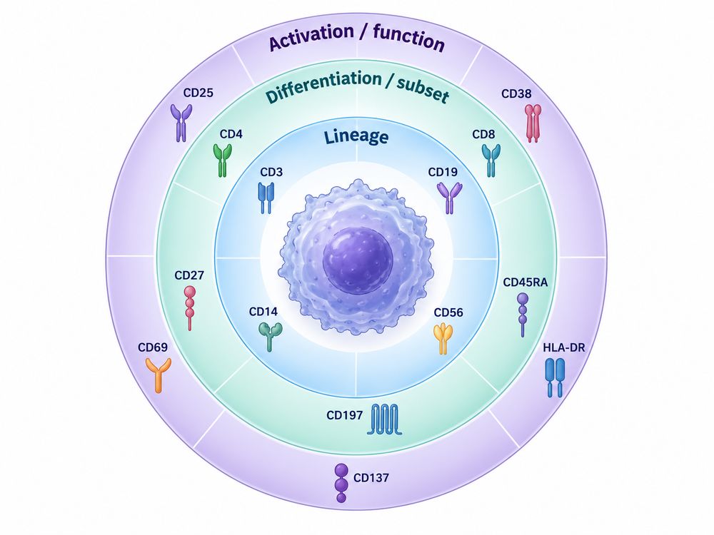 Figure 2. Three Layers of Immunophenotyping
This layered approach helps prevent a common panel-design error: allocating too many channels to activation markers before the population of interest can be robustly identified.
Common CD Marker Combinations
For practical CD markers flow cytometry workflows, combinations matter more than individual markers.
| Target Population | Example Marker Combination | Primary Purpose |
|---|---|---|
| Total T cells | CD3+ | T-cell lineage |
| Helper T cells | CD3+CD4+ | Major CD4 T-cell population |
| Cytotoxic T cells | CD3+CD8+ | Major CD8 T-cell population |
| Surface-defined Treg enrichment | CD3+CD4+CD25^high^CD127^low/−^ | Regulatory T-cell enrichment |
| B cells | CD3−CD19+ | B-cell identification |
| NK cells | CD3−CD56+ | NK-cell identification |
| Cytotoxic NK-rich population | CD3−CD56^dim^CD16+ | Major circulating NK subset |
| Classical monocytes | HLA-DR+CD14++CD16− | Classical monocyte population |
| Intermediate monocytes | HLA-DR+CD14++CD16+ | Intermediate monocyte population |
| Non-classical monocytes | HLA-DR+CD14+CD16++ | Non-classical monocyte population |
| Plasmacytoid DCs | Lin−HLA-DR+CD123+ | pDC enrichment |
| Conventional DC population | Lin−HLA-DR+CD11c+ | Broad conventional DC identification |
These examples should be treated as starting frameworks. Marker definitions can differ between laboratories, tissues, disease models, instruments, and analysis pipelines. Published OMIP and HIPC panels are valuable reference points because they document complete marker combinations and gating strategies rather than isolated markers. [2–4,7]
CD Markers in Multicolor and Spectral Flow Cytometry
Modern immunophenotyping increasingly measures many immune cell surface markers simultaneously. Conventional multicolor cytometry separates fluorescence using defined optical filters, whereas spectral flow cytometry collects broader emission information and computationally separates fluorochrome signatures.
Published full-spectrum panels have demonstrated simultaneous measurement of approximately 40 fluorescent parameters for deep immunophenotyping of human peripheral blood populations. [4]
Increasing the number of markers does not automatically improve an assay. High-dimensional panel development requires careful coordination of biology and optical performance.
Match Fluorochrome Brightness to Antigen Density
Dimly expressed markers generally benefit from brighter fluorochromes, while highly expressed lineage markers can often tolerate less bright channels. The optimal assignment also depends on instrument configuration and interactions among fluorophores.
Minimize Spillover and Spreading Error
Markers that must be resolved simultaneously should not be placed in fluorochrome combinations that generate excessive interference. Panel design is therefore best performed as a multidimensional optimization rather than selecting conjugates one marker at a time.
EuroFlow standardization work demonstrated that fluorochrome selection, cytometer configuration, instrument setup, compensation, sample preparation, antibody combinations, and analysis procedures all affect reproducibility. [6]
Use Appropriate Controls
Depending on the experiment, useful controls can include:
- unstained controls;
- single-stained compensation or spectral reference controls;
- fluorescence-minus-one controls;
- viability controls;
- biological positive and negative controls;
- antibody titration controls; and
- lot-to-lot bridging materials.
In spectral cytometry, accurate reference spectra are essential for reliable unmixing. Reference controls should reproduce the fluorophore behavior of experimental reagents as closely as practical.
Design the Biological Hierarchy First
A robust panel should normally prioritize:
- viability and singlet discrimination;
- broad leukocyte identification where required;
- lineage exclusion or inclusion;
- target population identification;
- subset differentiation;
- activation or functional markers.
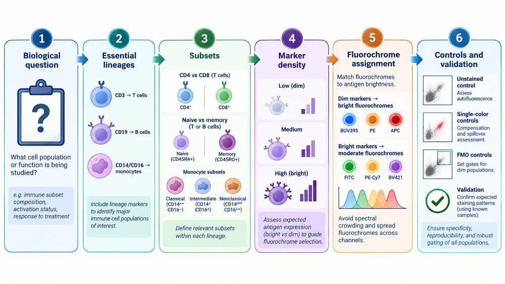 Figure 3. Building a Multicolor Immunophenotyping Panel
Explore Our CD Antigen Products
Learn More
CD Antigens as Controls and Standards in Assay Development
Recombinant CD proteins can support immunoassay and flow-related assay development, but their role should be distinguished from conventional cell-based controls.
Purified recombinant CD antigens may be useful for:
Antibody Binding and Specificity Testing
A purified extracellular CD domain provides a defined target for testing whether an antibody recognizes the intended antigen. This approach can complement positive and negative cell lines.
Reagent Lot Qualification
A stable antigen preparation can serve as part of a lot-bridging strategy to compare antibody binding between reagent lots.
Antibody Titration and Assay Optimization
Immobilized or bead-coupled antigen can help evaluate concentration-dependent binding and establish suitable assay conditions.
Receptor-Ligand and Fc Interaction Studies
Recombinant receptor ectodomains are useful for ELISA, SPR, BLI, and related binding formats. Fc receptor/CD16-related reagents are particularly relevant to studies of antibody-Fc receptor interactions.
Receptor-Expressing Cell Detection
Fluorophore-conjugated antigen can sometimes be used as a probe for cells engineered to express an antigen-specific receptor. For example, Creative BioMart describes a site-specific PE-labeled recombinant human CD4 reagent evaluated for staining anti-CD4 CAR-expressing cells.
Assay Standardization
Defined recombinant materials may contribute to long-term assay qualification when accompanied by appropriate purity, identity, activity, concentration, and lot documentation. Standardized immunophenotyping studies have shown more broadly that consistent reagents, instrument settings, staining procedures, and analysis methods substantially improve inter-laboratory comparability. [6,7]
However, recombinant proteins should not automatically be considered substitutes for cellular controls. A soluble ectodomain does not fully reproduce membrane orientation, receptor density, cellular microenvironment, post-translational modifications, or interactions with neighboring proteins.
The strongest validation strategy often combines defined molecular reagents with biologically relevant positive and negative cell controls.
How to Select Recombinant CD Antigens
The appropriate CD antigen reagents depend on the intended experimental application. Consider the following factors before selecting a recombinant protein.
| Selection Factor | Why It Matters | Practical Questions |
|---|---|---|
| Species | Antibodies and receptors may be species-specific | Human, mouse, cynomolgus, or another species? |
| Protein region | Epitopes may reside in specific extracellular domains | Full extracellular domain or defined fragment? |
| Expression system | Influences folding and post-translational modification | Mammalian, insect, E. coli, or cell-free? |
| Tag | Affects purification, immobilization, and detection | His, Fc, GST, Avi, or tag-free? |
| Biotinylation | Enables oriented capture on streptavidin surfaces | Random or site-specific biotinylation? |
| Biological activity | Important for functional binding studies | Has receptor/ligand binding been experimentally validated? |
| Purity | Impacts background and assay reproducibility | What analytical method and purity specification are provided? |
| Endotoxin | Particularly important in cell-based immune assays | Is low-endotoxin material required? |
| Label | May enable direct detection | Unlabeled, PE-, APC-, or other fluorophore-labeled? |
| Lot documentation | Supports reproducibility and regulated workflows | Is a COA and lot-specific QC available? |
1. Match the Construct to the Epitope
Before purchasing a recombinant CD antigen, confirm that the construct includes the epitope or receptor-binding region required for the assay. A short recombinant fragment may be appropriate for some antibodies but unsuitable for conformational epitopes.
2. Consider Native Folding and Glycosylation
Many CD proteins are glycoproteins containing disulfide bonds and structurally complex extracellular domains. Mammalian expression systems can therefore be preferable when native-like folding or glycosylation is important.
For simple antibody recognition of a linear epitope, a bacterial expression system may still be adequate.
3. Select the Tag According to the Assay Format
His-tagged proteins are convenient for purification and capture. Fc fusion proteins can improve stability or apparent avidity, but the Fc portion may complicate assays involving Fc receptors.
For experiments specifically involving CD16, CD32, CD64, or other Fc-binding molecules, tag selection deserves special attention because an Fc-tagged analyte can introduce unintended interactions.
4. Use Site-Specific Biotinylation When Orientation Matters
Avi-tagged, site-specifically biotinylated proteins can provide more consistent orientation on streptavidin-coated beads, biosensors, or plates than randomly modified proteins.
5. Look Beyond Purity
High purity does not necessarily mean that the recombinant protein is correctly folded or biologically active. For receptor-binding applications, activity data can be as important as SDS-PAGE purity.
A qualified CD antigen should therefore be selected according to the actual assay mechanism—not solely molecular weight, concentration, or price.
Explore Our CD Antigen Products
Learn More
Related CD Antigen Products
Creative BioMart provides recombinant proteins and related reagents targeting a range of immune-cell markers and Fc receptor-associated CD molecules. Examples identified in the current catalog include recombinant CD4, CD19, CD14, CD16/FCGR3A, CD38, and other immune-cell-associated proteins.
| Target | Immune Cell Relevance | Example Research Applications |
|---|---|---|
| CD4 | Major CD4 T-cell marker | Antibody validation, receptor-binding assays, CAR detection |
| CD19 | Major B-cell marker | Antibody binding, CAR-related assays, assay development |
| CD14 | Monocyte-associated marker | Antibody validation, monocyte-related research |
| CD16 / FCGR3A | NK cells, monocyte subsets; Fc receptor | Fc-receptor binding, antibody characterization, ADCC-related research |
| CD38 | Activation/differentiation marker | Immune activation and antibody-binding studies |
| CD68 | Macrophage-associated marker | Macrophage marker research |
For example, Creative BioMart currently lists active recombinant human CD14, recombinant human CD16, multiple FCGR3A/CD16a constructs, recombinant CD19 proteins, and recombinant CD4 proteins in different tag or labeling configurations.
Researchers selecting CD antigen resources should match the species, protein domain, expression system, tag, labeling strategy, and validated activity to the intended assay.
Key Takeaways
CD markers are the foundation of immune-cell immunophenotyping, but their greatest value comes from combinatorial interpretation.
CD3, CD19, CD56, CD14, and related lineage markers establish broad immune-cell identity. Markers such as CD4, CD8, CD16, CD27, CD38, CD45RA, CD69, CD123, and others then provide information about subsets, maturation, differentiation, or activation.
For multicolor and spectral flow cytometry, effective panel design requires more than choosing biologically relevant markers. Antigen density, fluorochrome brightness, spectral interactions, controls, sample preparation, instrument configuration, and standardized analysis all contribute to data quality. Multicenter standardization studies support the importance of controlling these variables for reproducible immunophenotyping. [6,7]
Finally, recombinant CD antigens can complement cell-based workflows by providing defined reagents for antibody characterization, binding analysis, reagent qualification, and assay development. Selecting the correct protein construct and validating its fitness for the intended application are essential for obtaining meaningful results.
Resource
-
From CD Markers to Immune Maps: A New Era in Immunology
-
The CD Map Is Still Growing: New CD Molecules and HLDA11
-
Antigen Density: Beyond CD-Positive
-
Spatial CD Biology: Why Location Matters in the Tumor Microenvironment
-
Logic-Gated CAR-T: Revolutionizing Cancer Therapy
-
Cluster of Differentiation
References
- Fernández-Calles J, Kužílková D, Hedin F, Bakardjieva-Mihaylova V, Škvárová Kramarzová K, van Zelm MC, et al.; Human Cell Differentiation Molecules (HCDM) organization. CD Molecules Nomenclature 2025: Antibody Validation and Expression Profiling of Immune System G Protein-Coupled Receptors. European Journal of Immunology. 2025;55(12):e70099. doi:10.1002/eji.70099.
- Moncunill G, Han H, Dobaño C, McElrath MJ, De Rosa SC. OMIP-024: Pan-leukocyte immunophenotypic characterization of PBMC subsets in human samples. Cytometry Part A. 2014;85(12):995–998. doi:10.1002/cyto.a.22580.
- Staser KW, Eades W, Choi J, Karpova D, DiPersio JF. OMIP-042: 21-color flow cytometry to comprehensively immunophenotype major lymphocyte and myeloid subsets in human peripheral blood. Cytometry Part A. 2018;93(2):186–189. doi:10.1002/cyto.a.23303.
- Park LM, et al. OMIP-069: Forty-Color Full Spectrum Flow Cytometry Panel for Deep Immunophenotyping of Major Cell Subsets in Human Peripheral Blood. Cytometry Part A. 2020;97(10):1044–1051. doi:10.1002/cyto.a.24213.
- Mair F, Prlic M. OMIP-044: 28-color immunophenotyping of the human dendritic cell compartment. Cytometry Part A. 2018;93(4):402–405. doi:10.1002/cyto.a.23331.
- Kalina T, Flores-Montero J, van der Velden VHJ, Martin-Ayuso M, Böttcher S, Ritgen M, et al. EuroFlow standardization of flow cytometer instrument settings and immunophenotyping protocols. Leukemia. 2012;26:1986–2010. doi:10.1038/leu.2012.122.
- Finak G, Langweiler M, Jaimes M, Malek M, Taghiyar J, Korin Y, et al. Standardizing Flow Cytometry Immunophenotyping Analysis from the Human ImmunoPhenotyping Consortium. Scientific Reports. 2016;6:20686.

Contact us or send an email at for project quotations and more detailed information.
Quick Links
-

Papers’ PMID to Obtain Coupon
Submit Now -

Refer Friends & New Lab Start-up Promotions