

T Cell CD Markers: Activation, Differentiation and Immune Profiling
Table of Contents
T cells are highly heterogeneous lymphocytes whose phenotype changes with lineage, activation state, differentiation history, tissue localization, and antigen exposure. For this reason, no single marker can fully define a T cell population. Instead, immune profiling usually relies on combinations of T cell CD markers, T-cell receptor-associated molecules, chemokine receptors, and functional markers.
At the most basic level, CD3 identifies conventional mature T cells, whereas CD4 and CD8 separate the major helper and cytotoxic compartments. Additional markers such as CD45RA, CD45RO, CD27, CD28, CD62L, CCR7, CD69, CD25, and CD71 provide information about differentiation and activation. Co-stimulatory and inhibitory receptors—including CD28, CD134, CD137, CD152, CD278, and CD279—add another layer of information about T-cell responsiveness and immune regulation.
Understanding how these markers relate to one another is important for flow cytometry, immune monitoring, antibody characterization, vaccine research, cancer immunology, cell therapy development, and other applications involving T cell CD antigen proteins.
Explore Our CD Antigen Products
Learn More
Major T Cell Lineage CD Markers
The T-cell receptor-CD3 complex is the defining antigen-recognition machinery of most mature T lymphocytes. The TCR recognizes antigen-derived peptides presented by major histocompatibility complex molecules, while the associated CD3 chains transmit intracellular activation signals.
For routine human immune profiling, CD3 is therefore one of the most useful pan-T-cell markers. CD3 is commonly combined with CD4 or CD8 to establish the major T-cell gates before additional activation, differentiation, or functional markers are evaluated.
CD2, CD5, and CD7 are also widely expressed by T cells and can support lineage characterization, although they are not absolutely specific to T cells. Their expression can additionally change in some hematological disorders, making multi-marker interpretation preferable to reliance on a single antigen.
| Marker | Major Relevance to T Cells | Typical Use in Immune Profiling | Important Consideration |
|---|---|---|---|
| CD3 | Component of the TCR signaling complex | Pan-T-cell identification | Highly useful for defining mature conventional T cells |
| CD2 | Adhesion and co-signaling molecule | Supporting T-cell lineage identification | Also expressed by NK cells and some other lymphocytes |
| CD5 | Modulates antigen receptor signaling | T-cell phenotyping and hematology panels | Also found on subsets of B cells |
| CD7 | Early T-lineage-associated molecule | T-cell and NK-cell profiling | Not T-cell specific |
| CD4 | MHC class II co-receptor | Identification of helper T-cell compartment | Also expressed by some non-T-cell populations |
| CD8 | MHC class I co-receptor | Identification of cytotoxic T-cell compartment | Can occur as CD8αα or CD8αβ depending on cell type |
| CD45 | Leukocyte common antigen | Leukocyte gating and differentiation analysis | Isoforms such as CD45RA and CD45RO provide additional information |
In practical flow cytometry, a basic sequence might begin with viable singlet leukocytes, followed by CD45-positive cells, CD3-positive T cells, and finally CD4/CD8 subdivision. The panel can then be expanded according to the biological question.
Researchers developing immunophenotyping or antibody-binding assays can use well-characterized CD antigen research proteins as assay reagents, reference materials, or targets during method development.
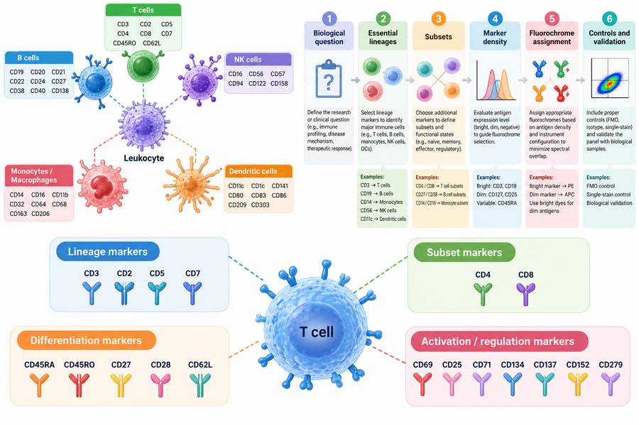 Fig 1. Major CD Markers Used to Define T Cell Identity and Functional State
CD4 vs CD8 T Cell Subsets
After identification of CD3-positive T cells, CD4 and CD8 are among the most informative markers for defining major T-cell populations.
CD4-positive T cells primarily recognize peptide antigens presented by MHC class II molecules. They include multiple functional populations, such as conventional helper T cells, regulatory T cells, follicular helper T cells, and other specialized states. Surface-marker combinations are required to distinguish these populations further.
CD8-positive T cells predominantly recognize peptide-MHC class I complexes. Following appropriate activation and differentiation, many acquire cytotoxic functions and can eliminate infected, malignant, or otherwise altered cells.
A simplified immune profiling hierarchy is:
CD45+ leukocytes → CD3+ T cells → CD4+ or CD8+ T cells → differentiation/activation subsets
However, CD4 and CD8 should not be interpreted as absolute synonyms for "helper" and "cytotoxic." Cytotoxic CD4 T-cell populations can occur, and CD8 T cells encompass multiple functional states rather than one homogeneous cytotoxic population.
Double-positive CD4+CD8+ and double-negative CD4−CD8− T cells can also be detected, although their frequency and biological significance depend strongly on age, tissue, disease state, and experimental setting.
For more detailed characterization, CD4/CD8 analysis is commonly combined with markers such as CD45RA, CCR7, CD27, CD28, CD25, CD127, CD38, HLA-DR, and PD-1.
Naive, Effector and Memory T Cell Markers
T cells change their surface phenotype substantially after antigen encounter. Among the most widely used approaches for human T-cell differentiation analysis is the combination of CD45RA and CCR7.
This framework separates circulating T cells into four commonly used phenotypic populations:
- Naive T cells: CD45RA+ CCR7+
- Central memory T cells (TCM): CD45RA− CCR7+
- Effector memory T cells (TEM): CD45RA− CCR7−
- Effector memory T cells re-expressing CD45RA (TEMRA): CD45RA+ CCR7−
CD45RO is frequently used as an additional memory-associated marker. CD27, CD28, CD62L, CD95, CD127, and other molecules can further resolve differentiation states.
| T Cell Population | Common Human Phenotype | Biological Characteristics |
|---|---|---|
| Naive T cells | CD45RA+ CCR7+; usually CD27+ CD28+ CD62L+ | Antigen-inexperienced cells with lymphoid homing and high proliferative potential |
| Central memory (TCM) | CD45RA− CCR7+; often CD45RO+ CD27+ | Memory cells with lymphoid homing and strong proliferative capacity |
| Effector memory (TEM) | CD45RA− CCR7−; generally CD45RO+ | More immediate effector potential and broader peripheral-tissue trafficking |
| TEMRA | CD45RA+ CCR7− | Highly differentiated population, particularly prominent within CD8 T cells |
| Stem cell-like memory (TSCM) | CD45RA+ CCR7+ CD95+ with additional naive-like markers | Rare antigen-experienced population with self-renewal and differentiation potential |
The CD45RA/CCR7 framework is extremely useful, but T-cell differentiation should not be regarded as a rigid series of completely discrete compartments. Modern studies increasingly describe antigen-experienced T cells as occupying a continuum of phenotypic and functional states. Marker definitions should therefore be explicitly reported rather than assuming that every laboratory uses identical gating criteria.
CD27 and CD28 are particularly useful as supplementary differentiation markers. Progressive differentiation—especially within some CD8 T-cell populations—can be accompanied by reduced expression of one or both molecules. CD57 and KLRG1 are sometimes included when studying highly differentiated T-cell populations, although their biological meaning depends on context.
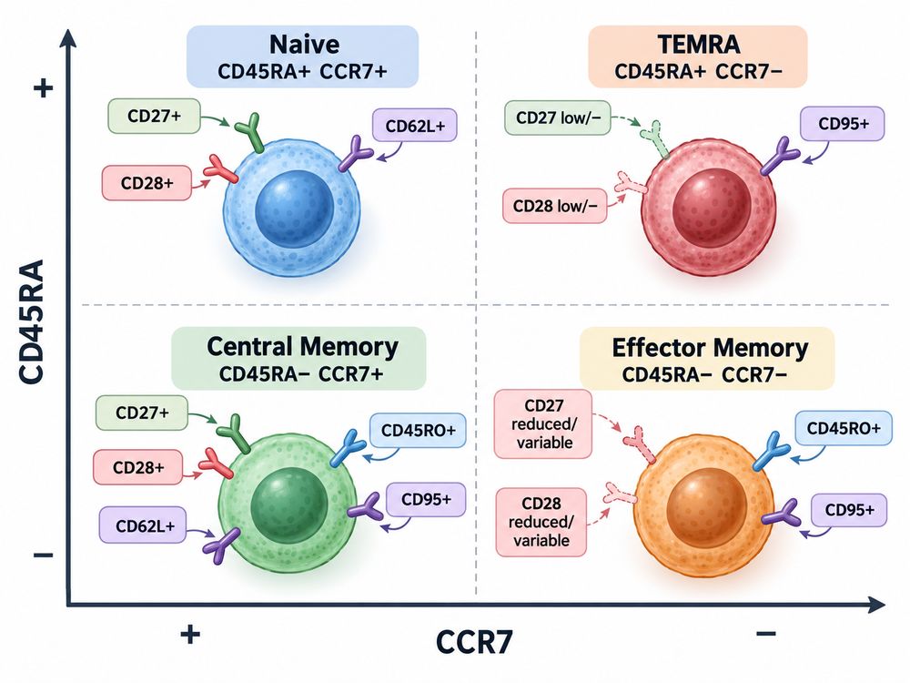 Fig 2. T Cell Differentiation Defined by CD45RA and CCR7
The selection of appropriate immune cell CD antigens is particularly important when building multicolor flow cytometry panels or validating antibodies intended to discriminate closely related T-cell populations.
Explore Our CD Antigen Products
Learn More
T Cell Activation Markers
T-cell activation is dynamic. TCR engagement initiates intracellular signaling within minutes, but different surface proteins appear at different stages and persist for different lengths of time.
Consequently, T cell activation markers should be selected according to the stimulation model and sampling time. CD69, CD25, and CD71 are commonly used, but each measures a somewhat different biological component of the activation response.
CD69
CD69 is one of the earliest surface proteins induced after T-cell activation and is therefore widely used as an early T cell activation marker.
Following TCR stimulation, CD69 can become detectable within hours. This makes it useful for short-term stimulation experiments, antigen-response assays, and rapid measurements of lymphocyte activation.
However, CD69 should not automatically be interpreted as evidence of recent antigen stimulation.
CD69 also has important roles in tissue retention and is frequently expressed by tissue-resident lymphocytes, including many tissue-resident memory T cells. In tissue samples, therefore, CD69 may reflect residency or localization biology as well as activation.
This distinction is important when comparing peripheral blood with tumor, mucosal, lung, skin, or other tissue-derived samples.
CD25
CD25 is the alpha chain of the high-affinity interleukin-2 receptor and is strongly induced on many T cells following activation.
Because IL-2 signaling supports T-cell proliferation and expansion, CD25 is widely used as a T cell activation marker, especially at later time points than the earliest CD69 response.
There is an important exception: regulatory T cells can constitutively express high levels of CD25. CD25 positivity within CD4 T cells should therefore not be interpreted automatically as recent activation.
For regulatory T-cell analysis, CD25 is commonly interpreted together with additional markers such as CD127 and intracellular FOXP3. For activation studies, CD25 is generally more informative when combined with other activation markers rather than used alone.
CD71
CD71 is the transferrin receptor 1 and mediates cellular iron uptake. Its expression increases when T cells undergo metabolic activation and proliferation.
Activated lymphocytes require substantial increases in biosynthetic activity to support cell growth and clonal expansion. Increased CD71 expression therefore provides a useful indicator of this metabolic and proliferative transition.
CD71 often complements CD69 and CD25:
- CD69 indicates relatively early activation.
- CD25 reflects development of an IL-2-responsive activation state.
- CD71 reflects increased metabolic demand associated with activated and proliferating cells.
CD71 is not specific to T cells and is expressed by many proliferating or metabolically active cell types. It should consequently be analyzed within a properly defined T-cell gate.
| Marker | Biological Meaning | Typical Interpretation | Major Limitation |
|---|---|---|---|
| CD69 | Rapidly induced activation-associated receptor | Early activation | Also associated with tissue residency |
| CD25 / IL-2Rα | High-affinity IL-2 receptor component | Activated/IL-2-responsive T cells | Constitutively high on many Tregs |
| CD71 / TfR1 | Iron uptake and metabolic activity | Metabolic activation and proliferation | Not specific to T cells |
| CD38 | Activation/metabolic-associated molecule | Often used with HLA-DR in immune monitoring | Expression varies by differentiation and disease state |
| HLA-DR | MHC class II molecule induced on activated T cells | Later or sustained activation | Not specific to T cells |
| CD154 / CD40L | Activation-induced ligand, especially on CD4 T cells | Functional antigen-response assays | Expression can be transient |
A robust activation panel therefore uses marker combinations plus an appropriate time point, rather than attempting to define activation using one molecule.
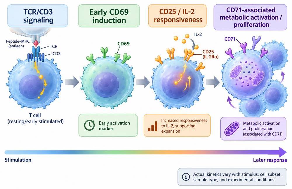 Fig 3. Dynamic Expression of T Cell Activation Markers
T Cell Co-stimulatory and Co-inhibitory Molecules
Antigen recognition through the TCR is only one component of T-cell activation. Co-signaling receptors determine whether TCR engagement is reinforced, sustained, modified, or inhibited.
Among the most important co-stimulatory molecules is CD28. Interaction of CD28 with CD80 or CD86 on antigen-presenting cells supports activation, cytokine production, survival, metabolic reprogramming, and proliferation.
Other co-stimulatory receptors can become particularly relevant after activation:
- CD27 interacts with CD70 and contributes to T-cell survival and memory responses.
- CD134/OX40 is induced after activation and can support expansion and survival.
- CD137/4-1BB is an activation-induced TNF receptor family member associated with enhanced T-cell survival and effector responses.
- CD278/ICOS provides co-stimulatory signaling, particularly in activated CD4 T cells and T follicular helper-cell biology.
Counterbalancing these pathways are inhibitory receptors.
CD152/CTLA-4 competes with CD28-associated pathways through interaction with CD80 and CD86 and is an important negative regulator of T-cell activation.
CD279/PD-1 interacts with PD-L1 and PD-L2 and can limit T-cell receptor signaling and effector activity. PD-1 expression is frequently evaluated in chronic antigen exposure, cancer, infection, and immune checkpoint research.
Other inhibitory receptors such as LAG-3/CD223, TIM-3, and TIGIT may also be incorporated into more specialized profiling panels.
Importantly, expression of an inhibitory receptor does not by itself prove T-cell exhaustion. PD-1, for example, can be transiently induced following normal activation. Functional state should be interpreted from combinations of phenotype, stimulation history, transcriptional state, cytokine production, proliferative capacity, and other measurements.
Recombinant receptor and ligand proteins—including CD marker proteins for immune research—can support studies of antibody specificity, receptor-ligand interactions, immune checkpoint biology, and functional assay development.
T Cell Marker Panels for Immune Profiling
There is no universal "best" T-cell marker panel. Panel design should start with the biological question.
A study intended only to quantify CD4 and CD8 T cells requires far fewer markers than an experiment designed to distinguish naive, memory, activated, regulatory, exhausted, and antigen-specific populations.
A practical strategy is to construct the panel in layers.
Basic T Cell Identification
A basic panel may include:
Viability → CD45 → CD3 → CD4 → CD8
This provides a foundation for measuring major circulating T-cell populations.
Differentiation Profiling
To study differentiation, add:
CD45RA + CCR7
Optional markers such as CD27, CD28, CD62L, CD95, or CD127 can increase resolution.
A typical differentiation panel might therefore include:
CD3 / CD4 / CD8 / CD45RA / CCR7 / CD27 / CD28
Activation Profiling
For short-term activation experiments:
CD3 / CD4 / CD8 / CD69 / CD25 / CD71
CD38 and HLA-DR may be added for studies of activated circulating T-cell populations, particularly when prolonged or systemic immune activation is relevant.
Regulatory T Cell Profiling
A commonly used regulatory T-cell strategy includes:
CD3 / CD4 / CD25 / CD127 / FOXP3
Because FOXP3 is intracellular, the workflow differs from an entirely surface-marker-based panel.
Co-signaling and Checkpoint Profiling
Depending on the research question, panels may include:
CD28 / CD27 / CD134 / CD137 / CD278 / CD152 / CD279 / CD223
The simultaneous measurement of activation and inhibitory receptors can be especially useful in cancer immunology, chronic infection, immunotherapy, and adoptive cell therapy research.
Practical Considerations for Panel Design
Marker selection is only one component of successful immune profiling. Researchers should also consider:
- Antigen density
- Fluorophore brightness
- Spectral overlap and compensation
- Autofluorescence
- Sample type
- Fresh versus cryopreserved samples
- Marker sensitivity to sample processing
- Stimulation duration
- Antibody clone
- Gating strategy
- Appropriate positive and negative controls
Some surface markers can change during sample processing or cryopreservation. Thus, assay conditions should be validated rather than assuming that a marker panel optimized for fresh peripheral blood will behave identically with frozen PBMCs or tissue-derived cells.
Current T-cell nomenclature recommendations also emphasize explicitly defining how populations were identified experimentally. This approach is preferable to using labels such as "effector," "memory," or "exhausted" without reporting the actual marker criteria.
Explore Our CD Antigen Products
Learn More
Recombinant CD Antigens for Antibody Validation and Functional Assays
Reliable immune profiling depends on reliable antibodies. Recombinant CD proteins can support antibody validation by providing a defined antigen independent of the biological variability associated with intact cells.
Depending on the protein format and assay design, recombinant CD antigens may be used in applications such as:
- Antibody binding and specificity testing
- Screening of antibody candidates
- ELISA development
- SPR or BLI binding analysis
- Receptor-ligand interaction studies
- Competition assays
- Assay positive controls
- Reference material development
- Immunization and antibody discovery workflows
- Cell-based assay development
- Immune checkpoint and co-stimulatory pathway research
Protein format is particularly important for cell-surface targets. Many CD molecules are membrane proteins whose native conformation depends on extracellular domain structure, oligomerization, glycosylation, disulfide formation, or membrane organization.
When choosing a recombinant antigen, researchers should therefore consider:
Species and sequence.
Human, mouse, non-human primate, and other orthologs may differ sufficiently to affect antibody binding.
Protein region.
For antibodies recognizing an extracellular epitope, an appropriately folded extracellular domain is generally more relevant than an unrelated intracellular fragment.
Expression system.
Mammalian expression may be preferred when native-like glycosylation or complex extracellular folding is important.
Tag configuration.
His, Fc, Avi, or other tags can simplify purification, immobilization, or assay development, but tag position may affect accessibility.
Biotinylation.
Site-specific biotinylated antigens can be advantageous for controlled immobilization in binding assays.
Purity and aggregation state.
Aggregated or heterogeneous proteins can produce misleading antibody-binding results.
Biological activity.
For receptor-ligand studies, biochemical purity alone may not demonstrate functional competence.
Using recombinant proteins alongside cell-based validation can provide complementary evidence. Protein-based assays establish direct recognition of a defined target, whereas cell-based assays help determine whether the antibody recognizes the antigen in its native cellular context.
Researchers working on T-cell phenotyping, antibody development, immune regulation, or receptor-ligand studies can explore additional recombinant CD antigen and immune receptor resources when selecting proteins for assay development and validation.
Ultimately, T-cell immune profiling is most informative when markers are interpreted as a coordinated phenotype rather than isolated labels. CD3, CD4, and CD8 define major lineage and subset relationships; CD45RA and CCR7 resolve important differentiation states; CD69, CD25, and CD71 provide complementary information about activation; and co-stimulatory and inhibitory receptors reveal additional regulatory states. Carefully selected marker combinations, validated reagents, and clearly defined gating criteria are therefore essential for reproducible T-cell analysis.
Explore Our CD Antigen Products
Learn More
Resource
-
From CD Markers to Immune Maps: A New Era in Immunology
-
The CD Map Is Still Growing: New CD Molecules and HLDA11
-
Antigen Density: Beyond CD-Positive
-
Spatial CD Biology: Why Location Matters in the Tumor Microenvironment
-
Logic-Gated CAR-T: Revolutionizing Cancer Therapy
-
Cluster of Differentiation

References
- Maecker HT, McCoy JP, Nussenblatt R. Standardizing immunophenotyping for the Human Immunology Project. Nature Reviews Immunology. 2012;12(3):191–200. doi:10.1038/nri3158.
- Cossarizza A, Chang HD, Radbruch A, et al. Guidelines for the use of flow cytometry and cell sorting in immunological studies (third edition). European Journal of Immunology. 2021;51(12):2708–3145. doi:10.1002/eji.202170126.
- Sallusto F, Lenig D, Förster R, Lipp M, Lanzavecchia A. Two subsets of memory T lymphocytes with distinct homing potentials and effector functions. Nature. 1999;401(6754):708–712. doi:10.1038/44385.
- Gattinoni L, Lugli E, Ji Y, et al. A human memory T cell subset with stem cell-like properties. Nature Medicine. 2011;17(10):1290–1297. doi:10.1038/nm.2446.
- Cibrián D, Sánchez-Madrid F. CD69: from activation marker to metabolic gatekeeper. European Journal of Immunology. 2017;47(6):946–953. doi:10.1002/eji.201646837.
- Poloni C, Schonhofer C, Ivison S, Levings MK, Steiner TS, Cook L. T-cell activation-induced marker assays in health and disease. Immunology & Cell Biology. 2023;101(6):491–503. doi:10.1111/imcb.12636.
- Rudd CE, Taylor A, Schneider H. CD28 and CTLA-4 coreceptor expression and signal transduction. Immunological Reviews. 2009;229(1):12–26. doi:10.1111/j.1600-065X.2009.00770.x.
- Masopust D, Awasthi A, Bosselut R, et al. Guidelines for T cell nomenclature. Nature Reviews Immunology. 2026;26:298–313. doi:10.1038/s41577-025-01238-2.

Contact us or send an email at for project quotations and more detailed information.
Quick Links
-

Papers’ PMID to Obtain Coupon
Submit Now -

Refer Friends & New Lab Start-up Promotions