

CD9, CD63 and CD81: Extracellular Vesicle and Exosome Markers
Table of Contents
- Why CD9, CD63 and CD81 Are Used in EV Research
- What Are Tetraspanins?
- CD9 in Extracellular Vesicles
- CD63 in Extracellular Vesicles
- CD81 in Extracellular Vesicles
- CD9 vs CD63 vs CD81
- Are They Universal Exosome Markers?
- Building an EV Characterization Panel
- EV Capture and Detection Assays
- Recombinant Tetraspanin Antigens
Why CD9, CD63 and CD81 Are Used in EV Research
CD9, CD63 and CD81 are among the most frequently measured proteins in extracellular-vesicle studies.
They belong to the tetraspanin family and can be abundant on the membranes of many EV populations.
As a result, antibodies against these CD antigens are widely used for:
- Western blotting;
- flow-based EV analysis;
- immunocapture;
- bead-based assays;
- single-vesicle detection.
However, their popularity can create an oversimplification.
CD9, CD63 and CD81 are not universally present on every EV, and none is completely specific to EVs.
Explore Our CD Antigen Products
Learn More
What Are Tetraspanins?
Tetraspanins are small membrane proteins containing four transmembrane domains.
They organize membrane microdomains and interact with other membrane proteins, lipids and cytoskeletal components.
The characteristic structure contains:
- four transmembrane helices;
- a small extracellular loop;
- a larger extracellular loop;
- short intracellular termini.
Tetraspanins participate in:
- adhesion;
- migration;
- signaling;
- membrane trafficking;
- vesicle formation.
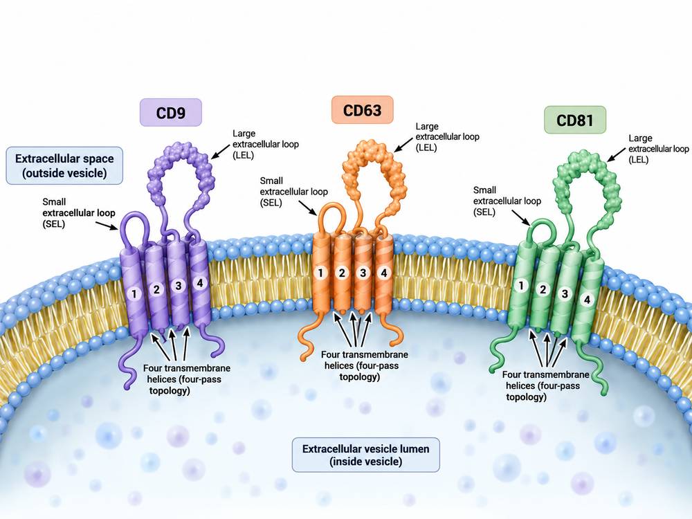 Fig 1. Tetraspanin Topology on an Extracellular Vesicle
CD9 in Extracellular Vesicles
CD9 is widely present on the plasma membrane and on many EV populations.
It is frequently detected in small EV preparations and is used in EV capture and detection assays.
However, CD9 abundance varies among cell types and vesicle subpopulations.
A CD9-negative particle is not necessarily non-vesicular.
CD63 in Extracellular Vesicles
CD63 is strongly associated with late endosomal and lysosomal compartments.
Because exosome biogenesis involves multivesicular endosomes, CD63 became one of the most widely recognized "exosome markers."
But CD63 also exists on cellular membranes and is not present uniformly in every exosome-like EV.
Therefore, CD63 positivity supports characterization but does not independently prove endosomal origin.
CD81 in Extracellular Vesicles
CD81 is another widely distributed tetraspanin.
It is involved in cell adhesion, signaling and membrane organization.
CD81 is frequently detected in EVs from many cell types and can complement CD9 and CD63 measurements.
CD9 vs CD63 vs CD81
| Marker | General EV Association | Key Limitation |
|---|---|---|
| CD9 | Common on many plasma-membrane-associated EV populations | Variable abundance |
| CD63 | Often enriched in endosomal compartments | Not universal or EV-specific |
| CD81 | Common EV tetraspanin | Cell-type dependent |
The relative expression of these three molecules can itself reveal EV heterogeneity.
A CD63-rich population may differ biologically from a CD9-rich population.
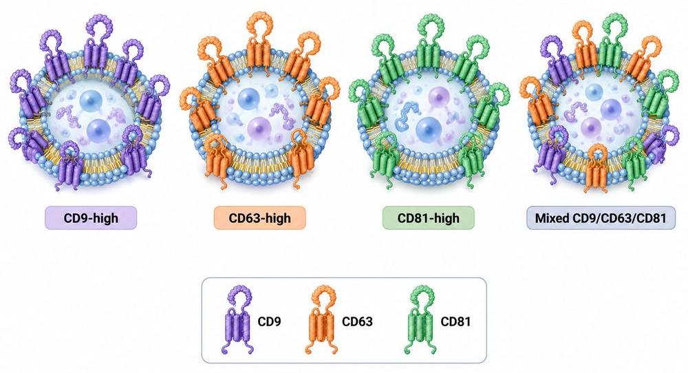 Fig 2. Heterogeneous EV Tetraspanin Profiles
Are They Universal Exosome Markers?
No.
MISEV2023 emphasizes that EV research requires multiple complementary characterization approaches.
The term "exosome" is best reserved for vesicles whose endosomal origin has been adequately demonstrated.
Detection of CD63 does not alone establish this origin.
Similarly, detection of CD9 or CD81 does not prove that every particle in an isolate is an EV.
Potential contaminants include:
- lipoproteins;
- protein aggregates;
- non-vesicular extracellular particles.
Therefore, researchers should avoid statements such as:
"CD63-positive particles are exosomes."
A more defensible conclusion is:
"The preparation contains extracellular particles positive for the EV-associated marker CD63."
Explore Our CD Antigen Products
Learn More
Building an EV Characterization Panel
A high-quality EV study usually combines multiple types of evidence.
EV-Associated Membrane Proteins
Examples include CD9, CD63 and CD81.
Cytosolic EV-Associated Proteins
These can provide evidence of vesicular cellular origin.
Contaminant Evaluation
Potential non-EV components should be assessed.
Particle Size and Concentration
Nanoparticle tracking analysis or related techniques can provide physical information.
Morphology
Electron microscopy or other imaging can provide structural evidence.
Single-EV Analysis
Advanced platforms can determine whether individual particles carry specific marker combinations.
| Method | Strength | Limitation |
|---|---|---|
| Western blot | Protein identity | Bulk measurement |
| NTA | Size/concentration | Limited phenotype information |
| TEM | Morphology | Limited quantitative phenotype |
| Bead flow cytometry | Multiplex surface markers | Indirect particle measurement |
| Single-EV assay | Resolves heterogeneity | Specialized technology |
EV Capture and Detection Assays
Tetraspanins are especially useful for immunocapture.
A common approach uses:
capture antibody → EV → detection antibody
For example:
anti-CD63 capture + anti-CD9 detection.
This design measures particles co-expressing both markers.
Changing the capture marker can change the population detected.
Therefore, comparing results from CD9-, CD63- and CD81-capture assays can reveal subpopulation differences.
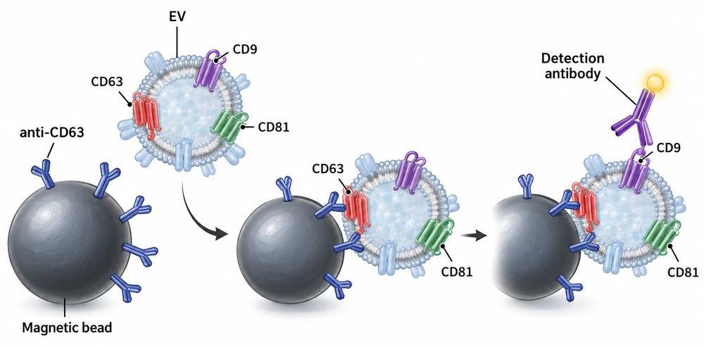 Fig 3. Tetraspanin-Based EV Immunocapture
Recombinant Tetraspanin Antigens
CD9, CD63 and CD81 are multipass membrane proteins.
That creates special challenges for recombinant expression.
A soluble extracellular loop may be sufficient for antibodies directed toward that loop, but it does not reproduce the complete membrane structure.
Therefore, validation should be matched to epitope type.
Recombinant CD antigen proteins can support:
- antibody screening;
- competition;
- standard curves;
- cross-reactivity testing.
But native EV or cell-based assays are important for antibodies recognizing membrane-dependent conformations.
For some applications, nanodisc- or membrane-based antigen presentation may provide better structural fidelity.
Are CD9, CD63 and CD81 exosome markers?
They are widely used EV-associated tetraspanin markers, but none is a universal or exclusive exosome marker. Reliable EV characterization should use multiple complementary markers and physical methods rather than defining an EV preparation from CD9, CD63 or CD81 alone.
Resource
-
From CD Markers to Immune Maps: A New Era in Immunology
-
The CD Map Is Still Growing: New CD Molecules and HLDA11
-
Antigen Density: Beyond CD-Positive
-
Spatial CD Biology: Why Location Matters in the Tumor Microenvironment
-
Logic-Gated CAR-T: Revolutionizing Cancer Therapy
-
Cluster of Differentiation

References
- Welsh JA, Goberdhan DCI, O'Driscoll L, et al. Minimal Information for Studies of Extracellular Vesicles (MISEV2023): From Basic to Advanced Approaches. J Extracell Vesicles. 2024;13(2):e12404. doi:10.1002/jev2.12404.
- Andreu Z, Yáñez-Mó M. Tetraspanins in Extracellular Vesicle Formation and Function. Front Immunol. 2014;5:442. doi:10.3389/fimmu.2014.00442.
- Kowal J, Arras G, Colombo M, et al. Proteomic Comparison Defines Novel Markers to Characterize Heterogeneous Populations of Extracellular Vesicle Subtypes. Proc Natl Acad Sci USA. 2016;113(8):E968–E977. doi:10.1073/pnas.1521230113.

Contact us or send an email at for project quotations and more detailed information.
Quick Links
-

Papers’ PMID to Obtain Coupon
Submit Now -

Refer Friends & New Lab Start-up Promotions