

Biotinylated Fc Receptor Proteins for SPR, BLI, and Screening Assays
Biotinylated Fc receptor proteins have become indispensable reagents in modern antibody characterization and therapeutic development. By leveraging site-specific biotinylation via Avi-tag technology, researchers can achieve uniform protein orientation on streptavidin-coated surfaces—critical for obtaining reproducible, high-quality binding data in Fc receptor interaction studies. This resource article explores the advantages, applications, and selection criteria for Avi-tagged Fc receptor proteins in SPR, BLI, and high-throughput screening workflows.
Table of Contents
- Why Use Biotinylated Fc Receptor Proteins?
- Advantages of Site-Specific Biotinylation
- Applications in SPR and BLI
- Applications in High-Throughput Antibody Screening
- Selecting Biotinylated FcγR, FcRn, FcεR, or FcαR Proteins
- Storage and Handling Considerations
- Creative BioMart Biotinylated Fc Receptor Protein Options
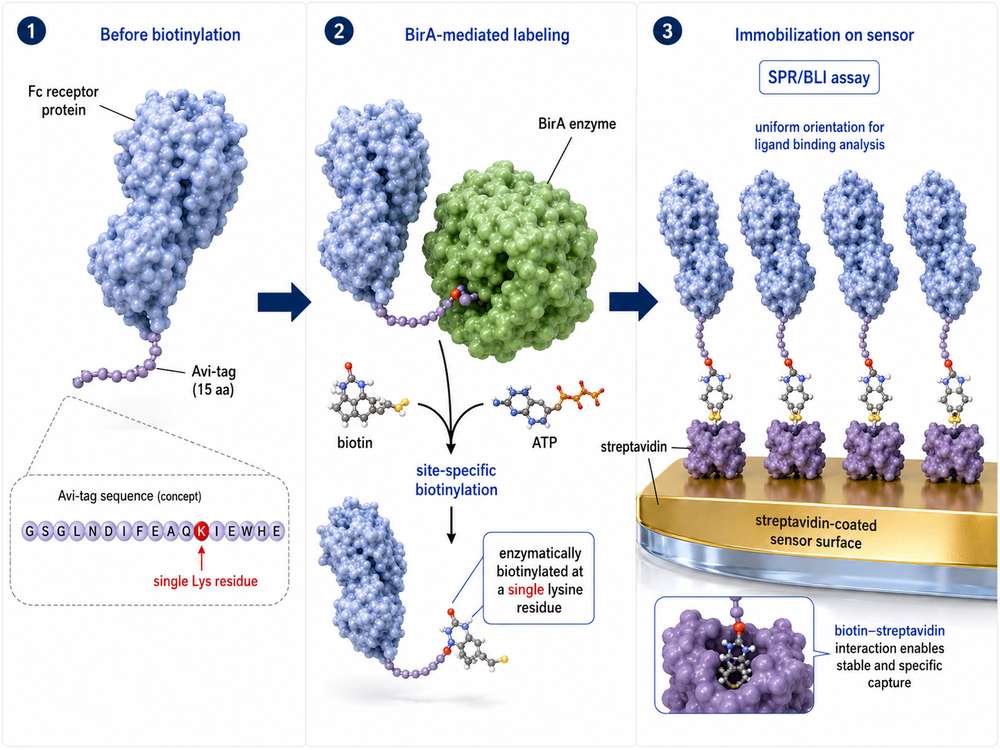 Fig1. Avi-Tag Site-Specific Biotinylation Workflow
Why Use Biotinylated Fc Receptor Proteins?
Fc receptors (FcRs) are critical mediators of antibody effector functions, binding to the Fc region of immunoglobulins to trigger downstream immune responses. In therapeutic antibody development, characterizing the interaction between candidate antibodies and various Fc receptors—including Fcγ receptors (FcγR), neonatal Fc receptor (FcRn), Fcε receptor (FcεR), and Fcα receptor (FcαR)—is essential for predicting efficacy, safety, and half-life.
Biotinylation provides a robust and versatile method for immobilizing Fc receptor proteins onto solid supports. The biotin-streptavidin interaction is one of the strongest non-covalent bonds known (affinity constant K ≈ 10¹⁴ M⁻¹), offering stable capture under diverse assay conditions. Compared to covalent coupling methods such as amine or thiol chemistry, biotin-based capture enables:
-
🔊 Reversible Immobilization
Streptavidin surfaces allow gentle regeneration without damaging the captured protein, enabling multiple binding cycles on the same sensor chip.
-
🔘 Controlled Orientation
Site-specific biotinylation at a defined location (via Avi-tag) ensures consistent protein orientation, preserving native binding epitopes.
-
⚡ High Sensitivity
The extraordinary affinity of biotin-streptavidin binding enables detection of weak or transient interactions with excellent signal-to-noise ratios.
-
🔀 Broad Compatibility
Biotinylated Fc receptors work seamlessly across SPR, BLI, ELISA, flow cytometry, and bead-based screening platforms.
Advantages of Site-Specific Biotinylation
Traditional chemical biotinylation modifies primary amines (lysine residues and N-termini) randomly across the protein surface. While straightforward, this approach can mask functional epitopes, alter protein conformation, and create heterogeneous labeling patterns that compromise assay reproducibility.
Avi-tag site-specific biotinylation overcomes these limitations through enzymatic precision. The Avi-tag is a unique 15-amino acid peptide (GLNDIFEAQKIEWHE) that contains a single biotin-acceptor lysine. The E. coli biotin ligase BirA recognizes this sequence and catalyzes the covalent attachment of biotin exclusively to that lysine residue.
Key Advantage: Because biotinylation occurs only on the Avi-tag lysine and not on the target protein itself, the natural binding activity of the Fc receptor remains fully preserved. When immobilized on an avidin-coated surface, the protein orientation is uniform because the biotin position is precisely controlled.
| Feature | Chemical Biotinylation | Avi-Tag Site-Specific Biotinylation |
|---|---|---|
| Labeling Site | Multiple random lysines / N-terminus | Single defined lysine in Avi-tag |
| Batch Consistency | Variable labeling density | Homogeneous, reproducible labeling |
| Impact on Activity | Risk of epitope masking | No interference with binding domains |
| Orientation Control | Random, mixed orientations | Uniform, directed orientation |
| Binding Kinetics | Variable, harder to interpret | Clean, interpretable sensorgrams |
| SPR/BLI Suitability | Moderate; may require optimization | Optimal; low coupling density possible |
| High-Throughput Screening | Higher assay variability | Excellent lot-to-lot consistency |
In Vivo vs. In Vitro Biotinylation
Avi-tag biotinylation can be performed through two primary routes:
- In vivo biotinylation: The Avi-tagged Fc receptor and BirA ligase are co-expressed in the host cells (e.g., HEK293 or CHO), with biotin supplied in the culture medium. This approach reduces post-expression processing steps and is ideal for large-scale production.
- In vitro biotinylation: Purified Avi-tagged protein is incubated with recombinant BirA, ATP, and biotin under controlled conditions. This method offers greater control over labeling efficiency and is preferred when >95% biotinylation is required for sensitive SPR/BLI applications.
For SPR and BLI assays where precise immobilization density directly impacts kinetic measurements, in vitro biotinylation often delivers more predictable and consistent performance.
Applications in SPR and BLI
Surface Plasmon Resonance (SPR) and Bio-Layer Interferometry (BLI) are label-free, real-time technologies used to quantify binding affinity, kinetics, and specificity between an immobilized ligand and a soluble analyte. Biotinylated Fc receptor proteins are particularly well-suited for these platforms.
SPR with Biotinylated Fc Receptors
In a typical SPR experiment, the biotinylated Fc receptor is captured at low density onto a streptavidin (SA) sensor chip. The antibody analyte is then injected at varying concentrations, and association/dissolution phases are monitored in real time. Site-specific biotinylation enables:
- Low-density, high-activity surfaces: Because every captured molecule is properly oriented, fewer molecules are needed to generate robust binding signals.
- Efficient regeneration: Gentle pH shifts or biotin competition can regenerate the streptavidin surface without denaturing the Fc receptor, allowing dozens of binding cycles per chip.
- Accurate kinetic modeling: Uniform orientation reduces mass transport artifacts and enables reliable 1:1 binding model fitting.
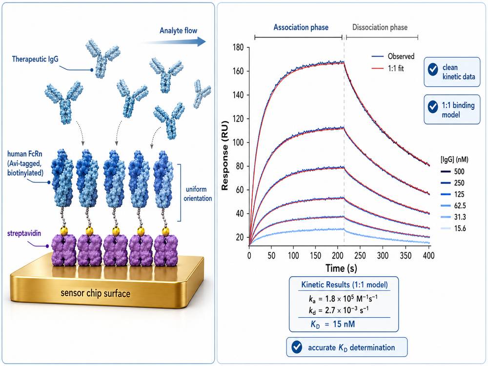 Fig 2. SPR Sensorgram of Biotinylated FcRn Binding to Therapeutic Antibody
BLI with Biotinylated Fc Receptors
BLI uses fiber-optic biosensors coated with streptavidin to capture biotinylated ligands. The dip-and-read format makes BLI especially attractive for:
- High-throughput kinetic screening: 96- or 384-well plate formats enable rapid profiling of multiple antibody candidates against a panel of Fc receptors.
- Crude sample analysis: BLI is more tolerant of complex matrices (e.g., cell culture supernatants) than SPR, streamlining early-stage screening.
- Parallel processing: Multiple sensors can be interrogated simultaneously, accelerating Fc receptor profiling during lead selection.
| Application | SPR | BLI |
|---|---|---|
| Affinity (KD) Determination | Gold standard; high precision | Excellent; plate-based throughput |
| Kinetic Rate Constants (kon/koff) | Superior resolution for fast kinetics | Good; suitable for most antibody-FcR interactions |
| Throughput | Low-to-moderate (serial flow) | High (parallel dip-and-read) |
| Sample Volume | ~50–200 µL per injection | ~200 µL per well |
| Regeneration | Multiple cycles per chip | Single-use disposable sensors |
| Best Use Case | Detailed characterization of lead candidates | Screening large antibody panels |
Applications in High-Throughput Antibody Screening
Therapeutic antibody development requires rapid evaluation of hundreds to thousands of candidates for their Fc receptor binding profiles. Biotinylated Fc receptor proteins enable streamlined, multiplexed screening across several assay formats:
Bead-Based Assays
Streptavidin-coated magnetic beads capture biotinylated Fc receptors, which then bind candidate antibodies from hybridoma supernatants or phage display libraries. Bound antibodies are detected via fluorescence or chemiluminescence, enabling rapid ranking by affinity.
ELISA and MSD Platforms
Biotinylated Fc receptors immobilized on streptavidin-coated plates serve as capture reagents in sandwich ELISAs. The uniform orientation maximizes epitope accessibility, improving assay sensitivity and dynamic range.
Flow Cytometry
Biotinylated Fc receptors can be coupled to fluorescent streptavidin conjugates for use in cell-based binding assays, enabling assessment of antibody binding in physiologically relevant contexts.
Phage Display and Biopanning
In antibody discovery, biotinylated Fc receptors are immobilized on streptavidin-coated surfaces to selectively enrich phage particles displaying FcR-binding antibody fragments, accelerating the identification of high-affinity clones.
Screening Tip: When screening large antibody panels, use a consistent biotinylated Fc receptor batch with confirmed >95% biotinylation efficiency (verified by HABA assay or mass spectrometry) to minimize batch-to-batch variability and ensure comparable ranking across plates.
Selecting Biotinylated FcγR, FcRn, FcεR, or FcαR Proteins
Choosing the right biotinylated Fc receptor format depends on your specific research objectives, assay platform, and therapeutic focus. Below is a selection guide organized by product characteristics:
| Selection Criterion | Considerations | Recommended Approach |
|---|---|---|
| Receptor Type | Effector function (ADCC/ADCP), half-life (FcRn), allergy (FcεR), mucosal immunity (FcαR) | Select Fc receptor panel matching therapeutic mechanism |
| Species | Human for clinical relevance; cynomolgus/rhesus for preclinical; mouse/rat for mechanistic studies | Match species to development stage |
| Allotype | FcγRIIIa F158 vs. V158 affects ADCC; FcγRIIa H131 vs. R131 affects phagocytosis | Include relevant allotypes for population coverage |
| Tag Configuration | Avi-tag position (N- vs. C-terminal), additional tags (His, Fc, Strep II) | C-terminal Avi-tag often preferred for extracellular domains |
| Biotinylation Method | In vivo vs. in vitro; efficiency verification | In vitro for SPR/BLI; in vivo acceptable for ELISA/screening |
| Expression System | HEK293 for native glycosylation; CHO for scalability; E. coli for non-glycosylated domains | HEK293 for FcγR and FcRn; E. coli for minimal domains |
| Validation Data | SPR/BLI verified, ELISA bioactivity, SDS-PAGE purity, endotoxin levels | Prioritize products with platform-specific validation |
Explore Our Fc Receptor Products
Learn more
Fcγ Receptor Panel Selection
For therapeutic antibodies targeting cancer or autoimmune diseases, a comprehensive FcγR panel should include:
- Activating receptors: FcγRI (CD64), FcγRIIa (CD32a, H131 allotype), FcγRIIIa (CD16a, V158 allotype)
- Inhibitory receptor: FcγRIIb (CD32b)
- Neonatal receptor: FcRn (heterodimer with β2-microglobulin) for half-life assessment
FcRn-Specific Considerations
FcRn binding is pH-dependent (strong at pH 6.0–6.5, weak at pH 7.4). When selecting biotinylated FcRn for SPR or BLI, ensure the assay buffer pH is carefully controlled, and consider using FcRn heterodimers (FcGRT + β2M) for physiologically relevant binding studies.
Storage and Handling Considerations
Proper handling of biotinylated Fc receptor proteins is essential to maintain biotin occupancy, protein stability, and biological activity. Follow these best practices:
| Parameter | Recommendation | Rationale |
|---|---|---|
| Storage Temperature | -20°C to -70°C (lyophilized); -70°C (reconstituted) | Minimizes protein degradation and biotin dissociation |
| Freeze-Thaw Cycles | Avoid repeated cycles | Each cycle risks aggregation and activity loss |
| Reconstitution | Sterile water or PBS; centrifuge before opening | Prevents contamination; recovers all lyophilized material |
| Working Aliquots | Prepare single-use aliquots at >100 µg/mL | Reduces freeze-thaw exposure; maintains concentration |
| Endotoxin | Use products with <1 EU/µg | Prevents immune cell activation in functional assays |
| Stability | Lyophilized: 12 months at -20°C; Reconstituted: 3 months at -70°C | Manufacturer-validated shelf life under optimal conditions |
| Buffer Compatibility | PBS pH 7.4, HEPES, or Tris-based buffers | Avoid extreme pH or reducing agents that may affect biotin-streptavidin binding |
Handling Tip: Before opening a lyophilized vial, briefly centrifuge at 4°C to ensure all powder settles at the bottom. Reconstitute with sterile water or buffer to the recommended concentration (typically 0.2–1 mg/mL), and avoid vortexing—gentle pipetting is sufficient.
Creative BioMart Biotinylated Fc Receptor Protein Options
Creative BioMart offers a comprehensive portfolio of biotinylated Fc receptor proteins produced using Avi-tag site-specific biotinylation technology. Our products are expressed in mammalian systems (HEK293 or CHO) to ensure proper folding, glycosylation, and biological activity.
Product Features
- Site-specific biotinylation: Single-point enzymatic labeling via Avi-tag ensures homogeneous product and uniform orientation
- High purity: >95% by SDS-PAGE; >90% by SEC-MALS
- Low endotoxin: <1.0 EU per µg by LAL method
- Verified bioactivity: Functionally tested by ELISA, SPR, and/or BLI
- Multiple species: Human, mouse, rat, cynomolgus, rhesus macaque
- Flexible tags: Avi-tag combined with His-tag, Fc-tag, or Strep II-tag
Available Formats
| Product Category | Tag Options | Expression System | Validation |
|---|---|---|---|
| Biotinylated FcγR Proteins (CD16a, CD32a, CD32b, CD64) |
His-Avi, Fc-Avi, Avi-His-Strep II | HEK293 | SPR, BLI, ELISA |
| Biotinylated FcRn Proteins (FcGRT & B2M heterodimer) |
His-Avi, Avi-Strep II | HEK293 | SPR, BLI |
| Biotinylated FcεR Proteins (FcεRI α-chain) |
His-Avi, Fc-Avi | HEK293 | ELISA, BLI |
| Biotinylated FcαR Proteins (CD89) |
His-Avi | HEK293 | ELISA |
| Custom Biotinylation Services | User-defined tag placement | HEK293 / CHO / E. coli | Custom QC per request |
Explore Our Full Fc Receptor Portfolio
Learn More
Custom Avi-Tag Biotinylation Services
For researchers requiring specialized Fc receptor constructs, Creative BioMart provides custom protein expression and site-specific biotinylation services. Our team can engineer Avi-tags at N-terminal, C-terminal, or internal loop positions, optimize BirA reaction conditions, and deliver proteins with defined biotin-to-protein ratios validated by HABA assay or mass spectrometry.
Resource
-
What Are Fc Receptors? The Immune System's Antibody Response Switch
-
FcRn: The Secret to Long-Lasting Antibodies!
-
ADCC vs ADCP: How Fcγ Receptors Drive Cancer Immunity
-
CD64: The Tiny Receptor with Big Implications!
-
Fc Receptor Polymorphisms: Why Antibody Responses Differ Between Patients
References
- Beck A, et al. (2010) Strategies and challenges for the next generation of therapeutic antibodies. Nature Reviews Immunology, 10(5): 345–352. doi:10.1038/nri2747
- Nimmerjahn F, Ravetch JV. (2008) Fcγ receptors as regulators of immune responses. Nature Reviews Immunology, 8(1): 34–47. doi:10.1038/nri2206
- Suzuki T, et al. (2010) Importance of neonatal FcR in regulating the serum half-life of therapeutic proteins containing the Fc domain of human IgG1: a comparative study of the affinity of serum albumin. Journal of Immunology, 184(4): 1968–1976. doi:10.4049/jimmunol.0902439
- Parrott MB, Barry MA. (2001) Metabolic biotinylation of secreted and cell surface proteins from mammalian cells. Biochemical and Biophysical Research Communications, 281(4): 993–1000. doi:10.1006/bbrc.2001.4443
- Howarth M, et al. (2006) A monovalent streptavidin with a single femtomolar biotin binding site. Nature Methods, 3(4): 267–273. doi:10.1038/nmeth861
- Schlothauer T, et al. (2013) Analytical FcRn affinity chromatography for functional characterization of monoclonal antibodies. mAbs, 5(4): 576–586. doi:10.4161/mabs.24981
- Jeong KJ, et al. (2017) AviTag-based detection and purification of complexes of a protein of interest and its binding partners. Methods in Molecular Biology, 1550: 243–258. doi:10.1007/978-1-4939-6745-2_16
- Li J, et al. (2019) High-throughput screening of antibody-Fc receptor interactions using biolayer interferometry. Journal of Pharmaceutical and Biomedical Analysis, 174: 297–305. doi:10.1016/j.jpba.2019.05.045
- Rodriguez ME, et al. (2013) The FcRn receptor: a key player in the development of biotherapeutics. Journal of Clinical Immunology, 33(Suppl 1): S1–S5. doi:10.1007/s10875-012-9795-4
- Gautier A, et al. (2008) An engineered protein tag for multiprotein labeling in living cells. Chemistry & Biology, 15(2): 128–136. doi:10.1016/j.chembiol.2008.01.007

Contact us or send an email at for project quotations and more detailed information.
Quick Links
-

Papers’ PMID to Obtain Coupon
Submit Now -

Refer Friends & New Lab Start-up Promotions