

FcγRIIb (CD32B) Proteins for Inhibitory Immune Signaling Research
FcγRIIb (also known as CD32B or FCGR2B) is the sole inhibitory member of the Fc receptor family expressed on human immune cells. Unlike activating FcγRs such as FcγRIIIA (CD16a), which drive antibody-dependent cellular cytotoxicity (ADCC) and pro-inflammatory responses, FcγRIIb functions as a critical immune checkpoint that maintains tolerance and prevents excessive immune activation. This resource provides a comprehensive overview of FcγRIIb biology, its role in antibody therapeutic safety and autoimmunity, and recombinant FCGR2B protein solutions for your research.
Table of Contents
- Overview of FcγRIIb / CD32B
- Inhibitory Fc Receptor Signaling and ITIM
- FcγRIIb in B Cells, Macrophages, and Mast Cells
- FcγRIIb and Antibody Therapeutic Safety
- FcγRIIb in Autoimmunity and Immune Regulation
- Binding Assay Considerations
- Recombinant FCGR2B Protein Formats
- Creative BioMart FCGR2B Solutions
Overview of FcγRIIb / CD32B
FcγRIIb (Fc fragment of IgG receptor IIb, gene symbol FCGR2B, also called CD32B) is a low-affinity receptor for the Fc region of IgG antibodies. Encoded by the FCGR2B gene on human chromosome 1q23, it is a 40 kDa type I transmembrane glycoprotein belonging to the immunoglobulin superfamily. FcγRIIb is unique among Fcγ receptors in that it is the only inhibitory receptor expressed on B lymphocytes and is co-expressed with activating Fc receptors on myeloid cells including macrophages, mast cells, dendritic cells, and neutrophils.
The extracellular domain of FcγRIIb contains two Ig-like domains (D1 and D2) that mediate binding to the Fc portion of IgG immune complexes. FcγRIIb binds all four human IgG subclasses (IgG1, IgG2, IgG3, IgG4) with low affinity, with a preference for IgG1 and IgG3. This low-affinity binding ensures that FcγRIIb is primarily engaged when immune complexes are present at high local concentrations, such as during secondary antibody responses or in the context of immune complex deposition in tissues.
Structurally, FcγRIIb exists as a single-chain receptor that does not require association with accessory signaling subunits (such as the FcRγ-chain or CD3ζ required by activating FcγRs). Instead, it contains an intrinsic immunoreceptor tyrosine-based inhibitory motif (ITIM) in its cytoplasmic tail, which is the molecular basis of its inhibitory function.
Key Distinction: While FcγRIIIA (CD16a) is an activating receptor that drives ADCC, ADCP, and cytokine release through ITAM-mediated SYK recruitment, FcγRIIb (CD32B) serves as the primary counter-regulatory mechanism, dampening these same effector functions via ITIM-mediated phosphatase recruitment.
Inhibitory Fc Receptor Signaling and ITIM
The inhibitory function of FcγRIIb is mediated through its cytoplasmic ITIM, a conserved six-amino-acid sequence (I/V/L/S)xYxx(L/V) that, upon phosphorylation, recruits phosphatases containing SH2 domains. The signaling cascade proceeds as follows:
- Co-ligation with activating receptors: When FcγRIIb is co-engaged with activating Fc receptors (e.g., FcγRIIIA on NK cells or B cell receptor on B cells) by IgG immune complexes, the ITIM is phosphorylated by Src-family kinases (Lyn, Fyn).
- Phosphatase recruitment: Phosphorylated ITIM serves as a docking site for SH2-domain-containing inositol 5-phosphatase (SHIP-1, also known as INPP5D) and SH2-domain-containing protein tyrosine phosphatases (SHP-1 and SHP-2).
- Signal termination: SHIP-1 hydrolyzes phosphatidylinositol (3,4,5)-trisphosphate [PIP3] to PIP2, thereby blocking the membrane recruitment and activation of downstream signaling molecules such as Akt, PLCγ, and Vav. SHP-1/SHP-2 directly dephosphorylate activating signaling intermediates.
- Biological outcome: The net result is suppression of calcium flux, inhibition of MAPK activation, reduced NF-κB signaling, and dampened cellular responses including degranulation, cytokine production, and proliferation.
This mechanism is not limited to FcγR signaling. FcγRIIb can also inhibit signaling from other activating receptors when co-expressed on the same cell, including B cell receptor (BCR), FcεRI on mast cells, and Toll-like receptors (TLRs) on dendritic cells and macrophages. This broad inhibitory capacity makes FcγRIIb a central regulator of immune homeostasis.
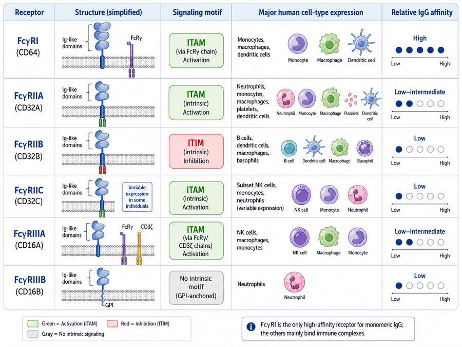 Fig1. (Left) Classification of human Fcγ receptors showing ITAM (green) and ITIM (red) motifs across FcγRI, FcγRII, FcγRIII families, with cell type expression patterns. (Right) FcγRIIb-mediated regulation of monoclonal antibody immunotherapy—direct targeting
 FcγRIIb in B Cells, Macrophages, and Mast Cells
The tissue distribution and functional impact of FcγRIIb vary significantly across immune cell types:
B Cells
On B lymphocytes, FcγRIIb is the only FcγR expressed. It plays a critical role in establishing the threshold for B cell activation and maintaining peripheral B cell tolerance. When the B cell receptor (BCR) and FcγRIIb are co-engaged by antigen-antibody immune complexes, FcγRIIb-mediated inhibition prevents excessive B cell proliferation, antibody production, and class switching. Mice deficient in Fcgr2b develop spontaneous lupus-like autoimmunity characterized by elevated autoantibody titers and glomerulonephritis, underscoring the essential role of FcγRIIb in preventing autoimmunity.
Macrophages
Macrophages express both activating FcγRs (FcγRI, FcγRIIIA) and the inhibitory FcγRIIb. The balance between activating and inhibitory signaling determines whether macrophages phagocytose antibody-opsonized targets (antibody-dependent cellular phagocytosis, ADCP) or release pro-inflammatory cytokines. FcγRIIb engagement on macrophages suppresses ADCP, reduces TNF-α and IL-6 production, and promotes an anti-inflammatory phenotype. In tumor microenvironments, tumor-associated macrophages (TAMs) often upregulate FcγRIIb, contributing to immune evasion by dampening ADCP of antibody-coated tumor cells.
Mast Cells
Mast cells express FcγRIIb alongside the high-affinity IgE receptor FcεRI. Co-engagement of FcγRIIb with FcεRI by IgG-containing immune complexes can suppress IgE-mediated degranulation and the release of histamine, leukotrienes, and pro-inflammatory cytokines. This mechanism is relevant to allergic diseases and may explain why certain IgG responses can attenuate IgE-mediated hypersensitivity reactions.
Table 1. Cell type-specific expression and functional roles of FcγRIIb (CD32B) in the immune system.
| Cell Type | FcγRIIb Expression Level | Primary Function | Clinical Relevance |
|---|---|---|---|
| B Cells | High (sole FcγR) | Sets activation threshold; maintains tolerance | Autoimmunity (SLE), antibody production regulation |
| Macrophages | Moderate to High | Suppresses ADCP and cytokine release | Cancer immune evasion, antibody therapy resistance |
| Mast Cells | Moderate | Inhibits IgE-mediated degranulation | Allergic diseases, anaphylaxis prevention |
| Dendritic Cells | Moderate | Modulates antigen presentation and T cell priming | Vaccine adjuvant design, tolerance induction |
| Neutrophils | Low to Moderate (inducible) | Limits NETosis and respiratory burst | Autoimmune neutrophil-mediated tissue damage |
FcγRIIb and Antibody Therapeutic Safety
The interaction between therapeutic monoclonal antibodies and FcγRIIb has emerged as a critical determinant of both efficacy and safety in antibody drug development:
FcγRIIb as a Barrier to Anticancer Antibody Efficacy
Many therapeutic antibodies used in oncology, including rituximab (anti-CD20), trastuzumab (anti-HER2), and cetuximab (anti-EGFR), rely on Fc-mediated effector functions (ADCC and ADCP) for their antitumor activity. However, FcγRIIb expressed on tumor-infiltrating immune cells (particularly macrophages and B cells within lymphoid tumors) can bind the Fc region of these therapeutic antibodies and deliver inhibitory signals that counteract activating FcγR signaling. This "inhibitory sink" effect can reduce the overall therapeutic potency of antibody drugs.
Preclinical studies have demonstrated that blocking FcγRIIb with antagonistic antibodies or engineering therapeutic antibodies with reduced FcγRIIb binding can enhance ADCC and ADCP, leading to improved tumor clearance. Conversely, increasing FcγRIIb affinity through Fc engineering has been explored as a strategy to enhance antibody internalization for antibody-drug conjugates (ADCs) or to improve B cell depletion in autoimmune indications.
FcγRIIb and Antibody-Dependent Enhancement (ADE)
In the context of infectious disease, particularly with dengue virus and SARS-CoV-2, FcγRIIb has been implicated in antibody-dependent enhancement (ADE) of infection. Sub-neutralizing antibody concentrations can form immune complexes that engage FcγRIIb on monocytes and macrophages, promoting viral uptake without triggering effective antiviral responses. Understanding the FcγRIIb binding profile of vaccine-induced or therapeutic antibodies is therefore essential for assessing the risk of ADE in infectious disease applications.
Safety Profile: Preventing Cytokine Release Syndrome
From a safety perspective, FcγRIIb engagement can be beneficial. By dampening macrophage and mast cell activation, FcγRIIb helps limit cytokine release syndrome (CRS) and infusion-related reactions associated with potent activating antibodies. Fc engineering strategies that selectively enhance FcγRIIb binding while preserving or reducing activating FcγR interactions represent an active area of research for improving the safety profile of immunotherapeutic antibodies.
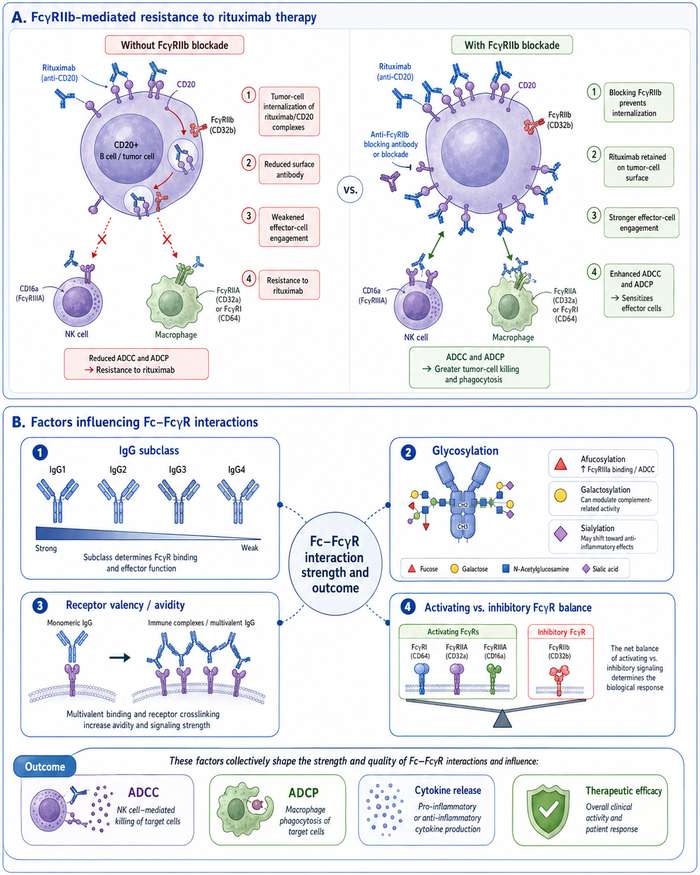 Fig2. (Top) FcγRIIb-mediated resistance to rituximab therapy—blocking FcγRIIb prevents tumor cell internalization and sensitizes effector cells. (Bottom) Factors influencing Fc-FcγR interactions, including IgG subclass, glycosylation, receptor valency, and activating vs. inhibitory FcγR engagement balance.
FcγRIIb in Autoimmunity and Immune Regulation
Genetic and functional evidence strongly supports a central role for FcγRIIb in the pathogenesis of autoimmune diseases:
Systemic Lupus Erythematosus (SLE)
The FCGR2B gene is located within the 1q23 locus, a region repeatedly identified in genome-wide association studies (GWAS) as associated with SLE susceptibility. A functional polymorphism in the transmembrane domain of FcγRIIb—Ile232Thr—impairs the receptor's ability to partition into lipid rafts and attenuates ITIM-mediated inhibitory signaling. Individuals carrying the Thr232 variant exhibit reduced FcγRIIb function, leading to:
- Lowered B cell activation thresholds and increased autoantibody production
- Enhanced immune complex-mediated inflammation in kidneys and skin
- Greater susceptibility to SLE, particularly in Asian populations where the Thr232 allele frequency is higher
Rheumatoid Arthritis and Other Autoimmune Diseases
Beyond SLE, FcγRIIb dysfunction has been linked to rheumatoid arthritis, idiopathic thrombocytopenic purpura (ITP), and autoimmune thyroiditis. In these conditions, impaired FcγRIIb signaling leads to insufficient suppression of immune complex-driven inflammation and tissue damage. Therapeutic strategies aimed at restoring or enhancing FcγRIIb function—such as IVIG (intravenous immunoglobulin) administration, which upregulates FcγRIIb expression on effector cells—have shown clinical benefit in multiple autoimmune indications.
Immune Tolerance and Allergy
FcγRIIb also contributes to the establishment of immune tolerance. During high-dose allergen immunotherapy, allergen-specific IgG antibodies form immune complexes that engage FcγRIIb on mast cells and basophils, suppressing IgE-mediated allergic responses. Similarly, FcγRIIb on dendritic cells can modulate T cell priming toward regulatory phenotypes, promoting tolerance rather than immunity.
Binding Assay Considerations
Accurate characterization of FcγRIIb-IgG interactions is essential for antibody engineering and drug development. Key assay considerations include:
Table 2. Key considerations for FcγRIIb binding assay design and execution.
| Assay Parameter | Consideration for FcγRIIb | Recommended Approach |
|---|---|---|
| Receptor Format | FcγRIIb is a single-chain receptor; no accessory subunits required | Use recombinant extracellular domain (ECD) proteins; full-length not necessary for binding |
| Binding Affinity | Low affinity (Kd ~10⁻⁶ M for IgG1 monomer); requires avidity for detection | Use multimeric IgG or immune complex formats; SPR with divalent analytes |
| Glycosylation | Fc glycan composition affects FcγRIIb binding (afucosylation reduces affinity) | Use glycoengineered or defined glycoform antibodies for consistent results |
| Polymorphism | Ile232Thr affects signaling but not IgG binding affinity | Use wild-type (Ile232) for binding assays; both variants for functional studies |
| Species Cross-Reactivity | Human and mouse FcγRIIb share ~60% homology; cynomolgus is closer to human | Use species-matched proteins for translational studies; cynomolgus for preclinical |
| Detection Method | Low affinity can yield weak signals in standard ELISA | Use BLI (Octet), SPR (Biacore), or AlphaLISA for sensitive, real-time detection |
SPR (Surface Plasmon Resonance) and BLI (Bio-Layer Interferometry) are the gold standard methods for measuring FcγRIIb-IgG binding kinetics. For these assays, biotinylated or His-tagged recombinant FcγRIIb protein can be immobilized on sensor chips, and the binding of IgG analytes (monomeric or multimeric) is monitored in real time. Because FcγRIIb binds monomeric IgG with low affinity, avidity effects from dimeric or multimeric IgG complexes often produce more robust signals.
Cell-based assays using FcγRIIb-expressing reporter cell lines (e.g., Jurkat-NFAT-CD32B) provide functional readouts of inhibitory signaling and are valuable for assessing the biological impact of Fc-engineered antibodies beyond simple binding affinity.
Recombinant FCGR2B Protein Formats
Recombinant FCGR2B proteins are indispensable tools for in vitro binding studies, antibody screening, and structural biology. The choice of protein format depends on the intended application:
Extracellular Domain (ECD) Proteins
The most commonly used format is the soluble extracellular domain (amino acids ~39–217 for human FcγRIIb), which encompasses both Ig-like domains responsible for IgG binding. ECD proteins are typically expressed in mammalian cells (HEK293 or CHO) to ensure proper folding and glycosylation, which are important for native-like IgG binding.
Tagged Variants
- His-tagged: Facilitates purification and immobilization on Ni-NTA surfaces for SPR/BLI assays.
- Avi-tagged + Biotinylated: Enables oriented immobilization on streptavidin-coated surfaces, improving assay reproducibility.
- GST-tagged: Useful for pull-down assays and as immunogens for antibody generation.
- Fc-tagged: Creates dimeric receptor formats that mimic natural avidity and enhance binding signals.
Species Variants
For translational research, recombinant FcγRIIb proteins from multiple species are available:
- Human: Primary target for therapeutic antibody development
- Cynomolgus monkey: Critical for preclinical safety and PK studies; shares ~95% homology with human FcγRIIb
- Mouse: Essential for in vivo mechanistic studies using mouse models
- Rat: Used in toxicology studies
Active vs. Non-Active Proteins
Some recombinant FcγRIIb preparations are functionally validated ("active") through binding assays (e.g., ELISA with human IgG1), confirming that the protein retains native IgG-binding activity. These are preferred for quantitative binding studies and drug discovery applications.
Creative BioMart FCGR2B Solutions
Creative BioMart offers a comprehensive portfolio of high-quality recombinant FCGR2B proteins and related Fc receptor research reagents to support your inhibitory immune signaling studies.
Table 3. Selected recombinant FCGR2B protein products available from Creative BioMart. Visit our FCGR2B product page for the complete catalog.
| Cat. No. | Product Name | Species | Source (Host) | Tag | Protein Length |
|---|---|---|---|---|---|
| FCGR2B-12820H | Recombinant Human FCGR2B, GST-tagged | E.coli | Human | GST | C-term-57a.a. |
| FCGR2B-2962H | Recombinant Human FCGR2B, His & AVI tagged | Human Cells | Human | Avi&His | 46-224 a.a. |
| Fcgr2b-3994M | Recombinant Mouse Fcgr2b, His & AVI tagged | Human Cells | Mouse | Avi&His | 1-217 a.a. |
| Fcgr2b-3995M | Recombinant Mouse Fcgr2b, His & AVI tagged | Human Cells | Mouse | Avi&His | |
| Fcgr2b-4083R | Recombinant Rat Fcgr2b protein(His 32 - Pro 212), His-tagged | HEK293 | Rat | His | His 32 - Pro 212 |
| FCGR2B-4543C | Active Recombinant Cynomolgus FCGR2B protein(Met1-Pro217), His-tagged | HEK293 | Cynomolgus | His | Met1-Pro217 |
| Fcgr2b-648M | Active Recombinant Mouse Fcgr2b protein(Met1-Arg217), His-tagged | HEK293 | Mouse | His | Met1-Arg217 |
| FCGR2B-650H | Active Recombinant Human FCGR2B Protein, His-tagged | HEK293 | Human | His | Ala46-Pro217 |
Quality Standards: All Creative BioMart recombinant FCGR2B proteins are manufactured under strict quality control with >95% purity (SDS-PAGE), endotoxin levels <1 EU/µg (LAL test), and bioactivity validation. Multiple expression systems (HEK293, CHO, E. coli, Human Cells) and tag options (His, GST, Avi, Fc, Biotinylated, Alexa Fluor conjugated) are available to match your experimental requirements.
Explore Our Complete Fc Receptor Research Portfolio
Learn More
FcγRIIb (CD32B) vs. FcγRIIIA (CD16a): Inhibitory vs. Activating Fc Receptors
To contextualize the unique role of FcγRIIb within the Fc receptor family, the following table provides a direct comparison with its activating counterpart, FcγRIIIA (CD16a):
Table 4. Side-by-side comparison of inhibitory FcγRIIB and activating FcγRIIIA. The balance between these receptors determines the net effect of therapeutic antibodies.
| Feature | FcγRIIB (CD32B) | FcγRIIIA (CD16a) |
|---|---|---|
| Gene Name | FCGR2B | FCGR3A |
| Receptor Type | Inhibitory (Single chain) | Activating (Requires FcRγ-chain) |
| Cytoplasmic Motif | ITIM (Immunoreceptor Tyrosine-based Inhibitory Motif) | ITAM (via FcRγ-chain adapter) |
| Signaling Outcome | Recruits SHP-1/SHP-2/SHIP → blocks activation | Recruits SYK → activates downstream cascades |
| Primary Cell Types | B cells, Macrophages, Mast cells, DCs, Neutrophils | NK cells, Macrophages, Monocytes |
| IgG Affinity | Low (binds IgG1, IgG2, IgG3 with low affinity) | Low to Intermediate (158V > 158F polymorphism) |
| Immune Function | Suppresses B cell activation, inhibits ADCC/ADCP, maintains tolerance | Mediates ADCC, ADCP, degranulation, cytokine release |
| Therapeutic Relevance | Antibody safety, autoimmunity regulation, immune tolerance | Cancer immunotherapy efficacy (e.g., trastuzumab, rituximab) |
| Clinical Polymorphism | Ile232Thr (affects ITIM signaling strength) | Val158Phe (affects IgG affinity & ADCC potency) |
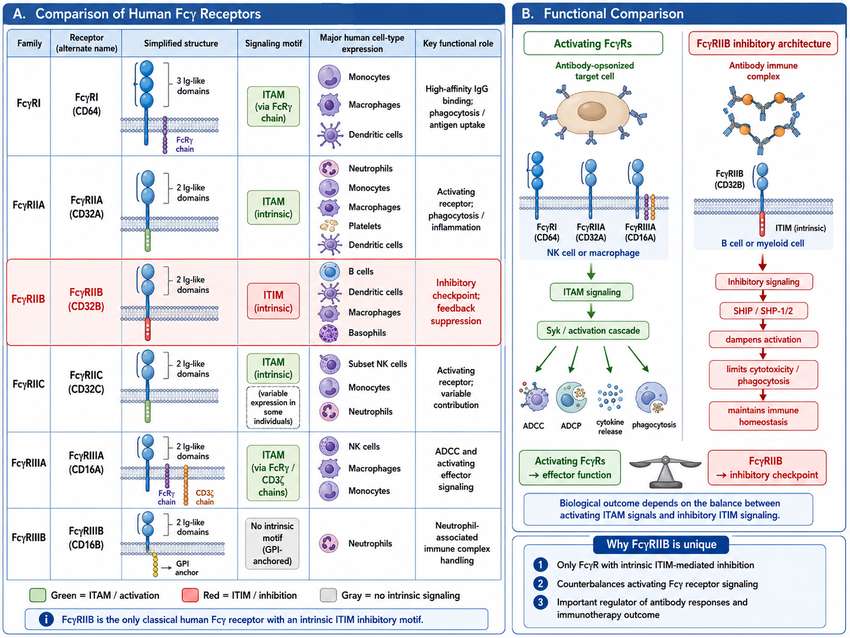 Fig 3. Structural and functional overview of human Fcγ receptors, illustrating the unique ITIM-containing inhibitory architecture of FcγRIIB compared to ITAM-bearing activating receptors.
Resource
-
What Are Fc Receptors? The Immune System's Antibody Response Switch
-
FcRn: The Secret to Long-Lasting Antibodies!
-
ADCC vs ADCP: How Fcγ Receptors Drive Cancer Immunity
-
CD64: The Tiny Receptor with Big Implications!
-
Fc Receptor Polymorphisms: Why Antibody Responses Differ Between Patients
References
- Kinet JP. The high-affinity IgE receptor (FcεRI): from physiology to pathology. Annu Rev Immunol. 1999;17:931-972.
- Gould HJ, Sutton BJ. IgE in allergy and asthma today. Nat Rev Immunol. 2008;8(3):205-217.
- Blank U, Rivera J. The ins and outs of IgE-dependent mast-cell exocytosis. Trends Immunol. 2004;25(5):266-273.
- Platzer B, Ruiter F, van der Molen R, et al. Soluble FcεRIα generates negative feedback activation of human basophils. J Allergy Clin Immunol. 2011;128(2):429-435.
- Dehlink E, Platzer B, Baker AH, et al. Solable FcεRIα: an endogenous immunoregulator in allergic disease? Clin Exp Allergy. 2011;41(7):943-951.
- Holgate ST, Polosa R. The mechanisms, diagnosis, and management of severe asthma in adults. Lancet. 2006;368(9537):780-793.
- Beck LA, Marcotte GV, MacGlashan D, et al. Omalizumab-induced reductions in mast cell FcεRI expression and function. J Allergy Clin Immunol. 2004;114(3):527-530.
- Davies AM, Allan EG, Keeble AH, et al. Allosteric mechanism of action of the therapeutic anti-IgE antibody omalizumab. J Biol Chem. 2017;292(24):9975-9987.
- Liu YJ, Wang HY, Wang R, et al. IgE-FcεRI protein-protein interaction as a therapeutic target against allergic asthma: An updated review. Int Immunopharmacol. 2025;139:113610.
- Harris G. Validating the trifunctional mechanism of action of Effector Cell Response Inhibitors (ECRIs). Presented at ACAAI 2025, Orlando, FL.
- Henningsson F, Yamamoto K, Saarinen JV, et al. Sialylation of IgE reduces FcεRIα interaction and mast cell and basophil activation in vitro and increases IgE half-life in vivo. Allergy. 2023;78(2):456-469.
- Greer AM, Wu N, Putnam AL, et al. Serum IgE clearance is facilitated by human FcεRI internalization. J Clin Invest. 2014;124(3):1187-1198.
- Tantisira KG, Lasky-Su J, Harada M, et al. Genomewide association between GLCCI1 and response to glucocorticoid therapy in asthma. N Engl J Med. 2011;365(13):1173-1183.
- Takai T. Fc receptors and their role in immune regulation and autoimmunity. J Clin Immunol. 2005;25(1):1-18.

Contact us or send an email at for project quotations and more detailed information.
Quick Links
-

Papers’ PMID to Obtain Coupon
Submit Now -

Refer Friends & New Lab Start-up Promotions