

FcRn Proteins for Antibody Half-Life and IgG Recycling Studies
Table of Contents
Explore Our Fc Receptor Products
Learn More
What Is FcRn?
The neonatal Fc receptor (FcRn) is a heterodimeric membrane glycoprotein that plays a central role in regulating the serum half-life of immunoglobulin G (IgG) antibodies and serum albumin. Structurally, FcRn consists of a major histocompatibility complex (MHC) class I-like α-chain encoded by the FCGRT gene non-covalently associated with β2-microglobulin (B2M). Unlike classical Fc receptors (FcγRs) that mediate effector functions such as antibody-dependent cellular cytotoxicity (ADCC), FcRn operates as a salvage receptor dedicated to intracellular trafficking, transcytosis, and protection of IgG from lysosomal degradation.
FcRn was first described in the 1960s as a receptor responsible for maternal IgG transport across the neonatal intestine. Subsequent research revealed that the same receptor also protects IgG from catabolism, establishing FcRn as a dual-function regulator of IgG homeostasis. In adults, FcRn is expressed in vascular endothelium, hematopoietic cells (macrophages, monocytes, dendritic cells), epithelial barriers, and placental syncytiotrophoblasts, where it maintains high circulating IgG levels essential for immune surveillance.
Key Takeaway: FcRn is structurally distinct from Fcγ receptors. Its primary function is not immune effector activation but rather pH-dependent IgG recycling and transcytosis, making it a critical determinant of antibody pharmacokinetics (PK).
FcRn, B2M, and IgG Binding
The functional FcRn receptor is a heterodimer comprising the FCGRT-encoded α-chain and B2M. The α-chain contains three extracellular domains (α1, α2, α3), a transmembrane region, and a cytoplasmic tail. The α1 and α2 domains form a peptide-binding groove structurally related to MHC class I molecules, while the α3 domain and B2M provide structural stability essential for proper folding and cell-surface expression.
IgG binds to FcRn at the interface between the CH2 and CH3 domains of the Fc region. Critical contact residues on the IgG Fc include Met252, Ile253, Ser254, Thr256, Thr307, His310, His435, and His436 (Eu numbering). On the FcRn side, key residues include His161, Ile166, and Tyr281, which form a hydrophobic pocket that accommodates the Fc CH2-CH3 hinge. The presence of B2M is indispensable for FcRn maturation and trafficking; B2M-deficient models exhibit severely impaired IgG recycling and dramatically reduced serum IgG levels.
Notably, FcRn binds monomeric IgG with 1:1 stoichiometry under physiological conditions, though recent evidence suggests that FcRn can engage both heavy chains of an IgG molecule in a 2:1 stoichiometry under certain conditions, which may influence the efficiency of recycling and the pharmacokinetic behavior of therapeutic antibodies.
pH-Dependent FcRn–IgG Interaction
The hallmark of FcRn function is its strict pH-dependent binding to IgG. At acidic pH (pH 5.5–6.5), characteristic of early endosomes, FcRn binds IgG with high affinity (KD ~0.3–0.8 µM for human IgG1). At neutral or physiological pH (pH 7.2–7.4), corresponding to the bloodstream and extracellular milieu, this interaction is negligible, allowing efficient release of recycled IgG back into circulation.
This pH switch is predominantly mediated by histidine residues that undergo protonation state changes across the physiological pH range:
- His310 (IgG Fc): Protonated at acidic pH, forms a salt bridge with FcRn Glu117, stabilizing the complex.
- His435 and His436 (IgG Fc): Critical for pH-dependent binding; mutation of His435 to Ala (H435A) abrogates FcRn interaction and results in extremely short antibody half-life.
- His161 (FcRn): The only residue on FcRn directly contributing to pH-dependent binding; its protonation facilitates interaction with IgG in acidic endosomes.
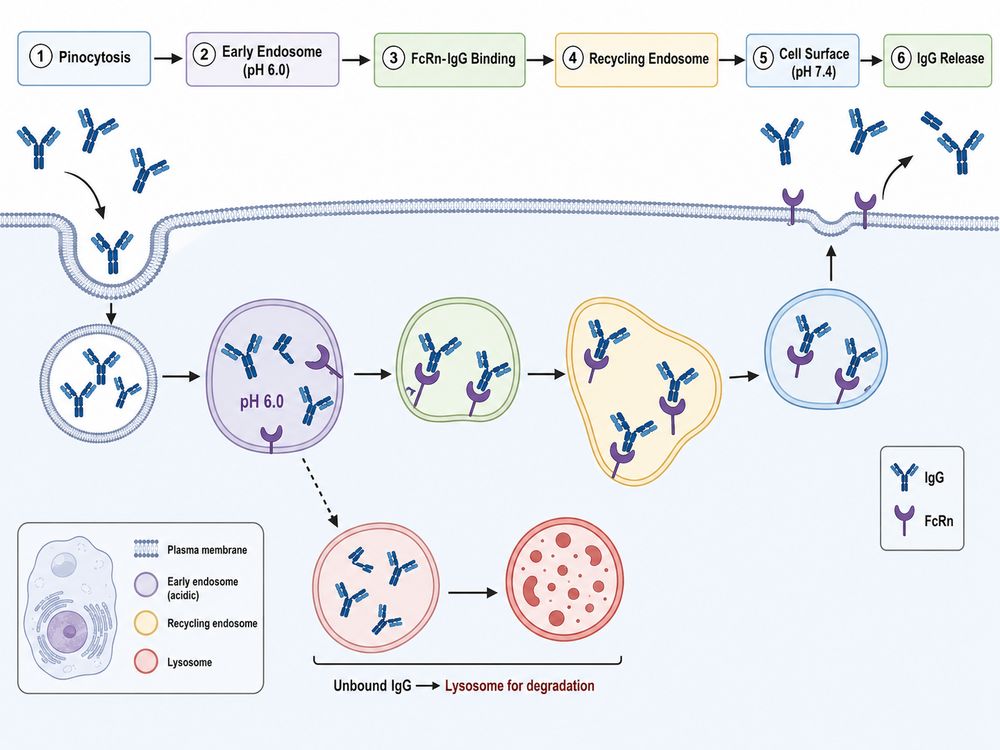 Fig 1. FcRn-mediated IgG recycling pathway. IgG is internalized via fluid-phase pinocytosis. In acidic endosomes, FcRn binds IgG and diverts it to recycling endosomes, bypassing lysosomal degradation. At the cell surface, neutral pH triggers IgG release.
The precise tuning of this pH threshold is essential for therapeutic antibody engineering. Variants that enhance FcRn binding at acidic pH while maintaining minimal interaction at neutral pH achieve the greatest half-life extension. Conversely, mutations that increase affinity at both pH values can lead to intracellular accumulation and impaired recycling, paradoxically shortening serum persistence.
FcRn in IgG Recycling and Serum Half-Life
FcRn-mediated recycling is the dominant mechanism governing the long serum half-life of IgG antibodies. In humans, IgG1, IgG2, and IgG4 exhibit half-lives of approximately 20–23 days, while IgG3—due to a single amino acid polymorphism at position 435 (Arg instead of His)—shows reduced FcRn binding and a shorter half-life of ~7 days.
The recycling process occurs primarily in vascular endothelial cells and hematopoietic cells through the following sequence:
- Fluid-phase pinocytosis: IgG is non-specifically internalized into early sorting endosomes.
- Acidification and binding: As endosomes mature and acidify to pH 6.0–6.5, FcRn binds IgG with high affinity.
- Sorting and recycling: The FcRn-IgG complex is sorted into recycling endosomes, diverting IgG away from lysosomal degradation pathways.
- Exocytosis and release: Upon fusion with the plasma membrane, the complex encounters neutral pH, triggering IgG dissociation and release into circulation.
When FcRn is saturated or when IgG lacks FcRn-binding capability, unbound antibodies are routed to lysosomes where they are degraded by proteases at acidic pH (≤5.5). This salvage mechanism is remarkably efficient: for every six albumin molecules recycled, approximately one IgG molecule is rescued in humans, yet this is sufficient to sustain high circulating IgG concentrations with minimal de novo synthesis.
Clinical Relevance: The FcRn recycling pathway is also the mechanistic basis for FcRn inhibitor therapies (e.g., efgartigimod, rozanolixizumab, nipocalimab). By blocking FcRn-IgG interaction, these agents accelerate the clearance of pathogenic autoantibodies in autoimmune diseases such as myasthenia gravis and immune thrombocytopenia.
FcRn in Therapeutic Antibody Engineering
Modulating FcRn binding affinity is one of the most validated strategies for extending the serum half-life of therapeutic antibodies and reducing dosing frequency. Two benchmark Fc engineering approaches have been clinically validated:
YTE Mutations (M252Y/S254T/T256E)
Originally developed at MedImmune/AstraZeneca, the YTE triple substitution enhances FcRn binding at pH 6.0 by approximately 10-fold through the creation of an additional salt bridge between Glu256 of Fc and Gln2 of B2M. YTE-engineered antibodies have demonstrated 2–4× half-life extension in humans. Clinical examples include:
- Beyfortus® (nirsevimab): Anti-RSV antibody with YTE mutations; approved for infant RSV prophylaxis with a single-dose regimen.
- Evusheld® (tixagevimab/cilgavimab): Anti-SARS-CoV-2 combination with YTE mutations; extended half-life up to ~100 days in adults.
LS Mutations (M428L/N434S)
Developed by Xencor, the LS double mutation increases hydrophobic interactions at the Fc-FcRn interface, enhancing pH 6.0 binding affinity. LS has been incorporated into:
- Ultomiris® (ravulizumab): Anti-C5 antibody for paroxysmal nocturnal hemoglobinuria (PNH); achieves extended dosing intervals.
- Sotrovimab: Anti-SARS-CoV-2 antibody with LS modifications.
Emerging Fc Engineering Strategies
Beyond YTE and LS, next-generation variants aim to optimize the kinetic balance between endosomal capture and serum release:
- DHS (L309D/Q311H/N434S): Exhibits 5-fold enhanced hFcRn binding at pH 5.8 with minimal neutral pH interaction, resulting in superior half-life extension compared to YTE/LS in transgenic mouse models.
- YML (L309Y/Q311M/M428L): A recently engineered variant demonstrating 6.1-fold half-life extension over wild-type in hFcRn transgenic mice, surpassing both DHS and benchmark variants.
- YD, DQ, DW variants: Novel combinations (M252Y/T256D; T256D/T307Q; T256D/T307W) showing comparable half-life extension to LS with improved developability profiles and reduced rheumatoid factor binding.
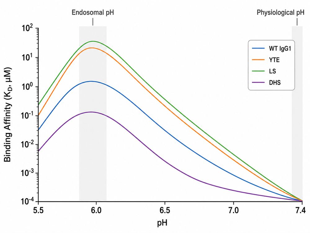 Fig 2. pH-dependent FcRn binding profiles of wild-type and engineered IgG Fc variants. The ideal therapeutic profile maximizes affinity at endosomal pH (~6.0) while ensuring complete dissociation at physiological pH (~7.4).
FcRn Binding Assays: SPR, BLI, ELISA
Quantitative characterization of FcRn-IgG interactions is essential for therapeutic antibody lead selection, optimization, and quality control. Three primary assay platforms are employed, each offering distinct advantages depending on the development stage and data requirements.
Table 1. Comparison of FcRn binding assay platforms for Antibody Development
| Assay Method | Principle | Kinetic Parameters | Throughput | pH Control | Best Application |
|---|---|---|---|---|---|
| SPR Surface Plasmon Resonance |
Label-free, real-time optical detection on gold sensor chip | ka, kd, KD; gold standard for affinity quantification | Medium (1–4 interactions/run) | Excellent (precise buffer exchange) | Lead optimization; detailed kinetic characterization; MOA studies |
| BLI Bio-Layer Interferometry |
Label-free, real-time interference-based detection at biosensor tip | ka, kd, KD; suitable for screening and ranking | High (96-well plate, parallel processing) | Good (buffer exchange between steps) | Early screening; clone ranking; formulation development |
| ELISA Cell-based / Recombinant |
Enzyme-labeled detection; endpoint or kinetic readout | EC50/IC50; relative affinity; limited kinetic resolution | Very High (384/1536-well compatible) | Moderate (static pH per well) | Batch release; stability testing; relative potency assays |
SPR (Surface Plasmon Resonance)
SPR remains the reference method for FcRn kinetic analysis. In a typical experiment, recombinant FcRn/FCGRT-B2M heterodimer is immobilized on a sensor chip (e.g., CM5 or CAP series), and IgG analytes are injected at pH 6.0 to measure association, followed by dissociation at pH 7.4. This configuration directly mimics the physiological pH transition and yields precise ka, kd, and KD values. SPR is particularly valuable for distinguishing variants with subtle differences in off-rate kinetics at neutral pH, a parameter increasingly recognized as critical for half-life optimization.
BLI (Bio-Layer Interferometry)
BLI offers a practical alternative for higher-throughput applications. Biosensor tips coated with FcRn are dipped into IgG-containing buffers at defined pH values, enabling parallel screening of multiple clones. While slightly less sensitive than SPR for very weak interactions, BLI excels in early-stage candidate ranking and formulation stability studies where speed and sample flexibility are prioritized.
ELISA-Based Approaches
ELISA formats for FcRn binding include direct binding assays (plate-coated FcRn with detection antibody) and competitive inhibition assays. Cell-based ELISA using FcRn-expressing cell lines (e.g., HMEC-1 endothelial cells) provides a more physiologically relevant context but introduces variability from endogenous protein expression. Recombinant FcRn ELISA offers better reproducibility for lot-release testing and biosimilar comparability studies.
Assay Design Consideration: When comparing FcRn binding across antibody variants, it is critical to maintain consistent pH conditions and to measure both association (pH 6.0) and dissociation (pH 7.4) phases. A variant with high affinity at pH 6.0 but slow dissociation at pH 7.4 may exhibit inferior in vivo half-life compared to a variant with moderate acidic pH affinity but rapid neutral pH release.
Species Considerations in FcRn Research
Species-specific differences in FcRn structure and IgG binding affinity have profound implications for preclinical pharmacokinetic studies and translational research. Selecting the appropriate model system is critical for accurate human PK prediction.
Table 2. Species-Specific FcRn Characteristics and Translational Relevance
| Species / Model | FcRn α-chain Homology | Binding to Human IgG1 | IgG half-life Prediction | Translational Value |
|---|---|---|---|---|
| Human | 100% | Reference (KD ~0.3–0.8 µM at pH 6.0) | ~21 days (IgG1/2/4) | Target species |
| Cynomolgus Monkey | ~95% | Comparable to human (~0.5–1.0 µM) | Good predictor; preferred NHP model | Gold standard preclinical PK; FIH dose selection |
| Mouse (WT) | ~65% | ~2.5× higher than human FcRn | Poor predictor; artificially prolonged PK | Not recommended for human PK extrapolation |
| Humanized Tg32 Mouse | human FCGRT transgene (endogenous promoter) | Human-equivalent affinity | Strong correlation (r² = 0.83 vs. human) | Cost-effective alternative to NHP; screening & optimization |
| Humanized Tg276 Mouse | Human FCGRT transgene (CAG promoter) | Human-equivalent (higher expression) | Strong correlation; higher FcRn expression | Mechanistic studies; may overestimate recycling |
Wild-Type Mouse Limitations
Wild-type mice bind human IgG1 with approximately 2.5-fold higher affinity than human FcRn, leading to artificially prolonged serum half-life and inaccurate human PK predictions. Additionally, endogenous murine IgG competes differently for FcRn binding, further complicating translational interpretation.
Humanized FcRn transgenic Models
The Tg32 and Tg276 mouse lines, in which the murine FCGRT gene is replaced with human FCGRT, have emerged as powerful alternatives to non-human primates:
- Tg32 mice express human FcRn under its endogenous promoter, recapitulating tissue-specific expression patterns similar to humans. PK clearance (CL) values from Tg32 mice correlate with human CL (r² = 0.83) even more strongly than cynomolgus monkey data (r² = 0.67), making them valuable for early screening.
- Tg276 mice drive human FcRn expression ubiquitously via the CAG promoter, resulting in higher overall receptor levels. While useful for mechanistic studies, this model may overestimate recycling capacity compared to physiological human expression.
Allometric scaling from Tg32 mice to cynomolgus monkeys (and by extension to humans) uses a clearance exponent of ~0.91, enabling quantitative translation of preclinical PK data. These models are particularly valuable for differentiating Fc-engineered variants during lead optimization, potentially reducing NHP usage in early development.
Creative BioMart FcRn and FCGRT/B2M Protein Solutions
Creative BioMart provides a comprehensive portfolio of recombinant FcRn proteins, FCGRT-B2M heterodimers, and related assay reagents designed to support antibody half-life studies, PK modeling, and FcRn binding assay development.
Recombinant FcRn Protein Products
- Human FcRn / FCGRT & B2M Heterodimer: Fully assembled heterodimeric receptor produced in mammalian cells (HEK293), ensuring correct glycosylation and conformation. Available with AviTag™, His Tag, and Strep II Tag for flexible assay integration.
- Species-Specific FcRn Variants: Recombinant cynomolgus monkey, mouse, rat, and rabbit FcRn proteins for cross-species comparative studies and translational PK research.
- FCGRT and B2M Monomers: Individual α-chain (FCGRT) and β2-microglobulin (B2M) subunits for reconstitution studies, structural biology, and custom heterodimer assembly.
Quality Verification and Bioactivity
All FcRn proteins are rigorously characterized to ensure batch-to-batch consistency and functional integrity:
- Purity: >95% by SDS-PAGE; >90% by SEC-HPLC
- SPR Verified: Binding affinity to human IgG1 Fc confirmed by Biacore (KD ~0.37–0.73 µM at pH 6.0)
- BLI Verified: Octet-based kinetic characterization for high-throughput compatibility
- ELISA Verified: Functional binding to therapeutic antibodies (e.g., trastuzumab, bevacizumab) demonstrated in plate-based assays
Applications in Antibody Development
Creative BioMart FcRn proteins support critical workflows across the therapeutic antibody development pipeline:
- Lead Selection & Optimization: Rank candidate antibodies by FcRn binding affinity using SPR/BLI with recombinant human FcRn as the capture molecule.
- Biosimilar Characterization: Demonstrate analytical similarity between innovator and biosimilar products through comparable FcRn binding profiles.
- Stability & Forced Degradation: Monitor FcRn binding as a quality attribute during accelerated stability studies and formulation development.
- FcRn Inhibitor Screening: Evaluate small molecules, peptides, or antibodies that disrupt FcRn-IgG interaction for autoimmune disease drug discovery.
Explore FcRn Protein
Learn More
Resource
-
What Are Fc Receptors? The Immune System's Antibody Response Switch
-
FcRn: The Secret to Long-Lasting Antibodies!
-
ADCC vs ADCP: How Fcγ Receptors Drive Cancer Immunity
-
CD64: The Tiny Receptor with Big Implications!
-
Fc Receptor Polymorphisms: Why Antibody Responses Differ Between Patients
References
- Roopenian DC, Akilesh S. FcRn: the neonatal Fc receptor comes of age. Nat Rev Immunol. 2007;7(9):715-725.
- Chaudhury C, et al. The major histocompatibility complex-related Fc receptor for IgG (FcRn) binds albumin and prolongs its lifespan. J Exp Med. 2003;197(3):315-322.
- Martin WL, Bjorkman PJ. Characterization of the 2:1 complex between the class I MHC-related Fc receptor and its Fc ligand in solution. Biochemistry. 1999;38(39):12639-12647.
- Dall'Acqua WF, et al. Increasing the affinity of a human IgG1 for the neonatal Fc receptor: biological consequences. J Immunol. 2002;169(9):5171-5180.
- Zalevsky J, et al. Enhanced antibody half-life improves in vivo activity. Nat Biotechnol. 2010;28(2):157-159.
- Yeung YA, et al. Engineering human IgG1 affinity to human neonatal Fc receptor: impact of affinity improvement on pharmacokinetics in primates. J Immunol. 2009;182(12):7663-7671.
- Deng R, et al. Pharmacokinetics of humanized monoclonal anti-tumor necrosis factor-alpha antibodies in simulated innate immune humanized mice. MAbs. 2010;2(5):515-521.
- Lee CH, et al. An engineered human Fc domain that behaves like a pH-toggle switch for ultra-long circulation persistence. Nat Commun. 2019;10:5031.
- Schlothauer T, et al. Novel human IgG1 and IgG4 Fc-engineered antibodies with completely abolished immune effector functions. Protein Eng Des Sel. 2016;29(10):457-466.
- Avery LB, et al. Establishing in vitro-in vivo correlation using human FcRn transgenic mice for predicting human pharmacokinetics of monoclonal antibodies. mAbs. 2018;10(2):246-255.
- Leabman MK, et al. Effects of altered FcRn binding affinity on the pharmacokinetics of monoclonal antibodies in cynomolgus monkeys. mAbs. 2013;5(5):778-786.
- Petkova SB, et al. Enhanced half-life of genetically engineered human IgG1 antibodies in a humanized FcRn mouse model: potential application in humorally mediated autoimmune disease. Int Immunol. 2006;18(12):1759-1769.
- Strohl WR. Fusion proteins for half-life extension of biologics as a strategy to make biobetters. BioDrugs. 2015;29(4):215-239.
- Grevys A, et al. Fc engineering for enhanced serum half-life. In: Antibody Fc. Academic Press; 2023:245-268.
- Schneider-Merck T, et al. Human IgG2 antibodies against epidermal growth factor receptor effectively trigger antibody-dependent cellular cytotoxicity but, in contrast to IgG1, only by cells of myeloid lineage. J Immunol. 2010;184(1):512-520.
- Borvak J, et al. Functional mapping of the FcRn-IgG interaction. J Immunol. 1998;161(12):6834-6841.
- Oganesyan V, et al. Structural characterization of a human Fc fragment engineered for lack of effector functions. Acta Crystallogr D Biol Crystallogr. 2008;64(Pt 7):700-704.
- Hinton PR, et al. Engineered human IgG antibodies with longer serum half-lives in primates. J Biol Chem. 2004;279(8):6213-6216.
- Monnet C, et al. Effector function rescue of a non-lytic bispecific antibody by immune cell-mediated killing. MAbs. 2020;12(1):1740103.
- Vaccaro C, et al. Engineering the Fc region of immunoglobulin G to modulate in vivo antibody levels. Nat Biotechnol. 2005;23(10):1283-1288.

Contact us or send an email at for project quotations and more detailed information.
Quick Links
-

Papers’ PMID to Obtain Coupon
Submit Now -

Refer Friends & New Lab Start-up Promotions