

Fcα Receptor and CD89 Proteins for IgA-Mediated Immunity
Quick Navigation
What Is FcαRI/CD89?
FcαRI, also known as CD89 or the FCAR protein, is the only canonical Fc receptor specific for immunoglobulin A (IgA). It is a type I transmembrane glycoprotein belonging to the immunoglobulin superfamily, composed of two extracellular Ig-like domains (EC1 and EC2), a transmembrane region, and a short cytoplasmic tail. Unlike many other Fc receptors, FcαRI lacks intrinsic signaling motifs and requires association with the Fc receptor gamma chain (FcRγ) to mediate intracellular signaling.
The human CD89 protein is encoded by the FCAR gene located on chromosome 19. Structurally, each IgA1-Fc molecule can interact with two FcαRI molecules at the Cα2–Cα3 junction, potentially inducing receptor dimerization on the cell membrane. This unique binding architecture distinguishes FcαRI from IgG and IgE receptors, which typically form 1:1 complexes with their ligands. The crystal structure of the FcαRI-IgA1-Fc complex, first resolved in 2003, revealed critical insights into the molecular basis of IgA-mediated immune recognition.
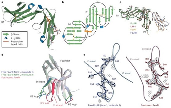 Fig 1. Crystal structure of human FcαRI (CD89) in complex with IgA1-Fc. The receptor binds at the Cα2–Cα3 junction of IgA, with two FcαRI molecules interacting with each IgA Fc dimer. (Source: Herr et al., Nature 2003)
Key Structural Features of CD89/FcαRI:
- Two extracellular Ig-like domains (EC1/EC2) oriented at approximately right angles
- Transmembrane arginine 209 (R209) essential for FcRγ-chain association
- Short cytoplasmic tail lacking intrinsic signaling motifs
- Binding site on IgA-Fc overlaps with polymeric Ig receptor (pIgR) binding site
Fcα Receptors and IgA Immune Responses
The Fc alpha receptor system operates through a sophisticated dual-signaling mechanism that enables IgA to function as both an anti-inflammatory and pro-inflammatory mediator. This functional duality is central to IgA-mediated immunity and depends critically on the oligomeric state of IgA and the extent of FcαRI crosslinking.
Monomeric serum IgA (non-complexed) binds to FcαRI without inducing receptor crosslinking. This interaction triggers inhibitory ITAM (ITAMi) signaling through the associated FcRγ-chain. The mechanism involves partial phosphorylation of ITAMs, recruitment of the tyrosine phosphatase SHP-1, and formation of intracellular "inhibisome" clusters that suppress phosphorylation of Syk, LAT, and ERK. Consequently, monomeric IgA dampens pro-inflammatory responses initiated by other activating Fc receptors, such as IgG-mediated FcγR activation.
In contrast, IgA immune complexes (e.g., IgA-opsonized pathogens) induce robust FcαRI crosslinking, leading to full ITAM phosphorylation by Src kinases (Lyn/Fyn). This creates docking sites for Syk and downstream signaling molecules including PI3K, PLCγ, and Shc, ultimately activating the Ras/Raf/MEK/MAPK pathway and NF-κB. The outcome is potent pro-inflammatory effector functions: phagocytosis, antibody-dependent cellular cytotoxicity (ADCC), respiratory burst, degranulation, cytokine release, and neutrophil extracellular trap (NET) formation.
| IgA Form | FcαRI Engagement | Signaling Pathway | Functional Outcome |
|---|---|---|---|
| Monomeric serum IgA | Non-crosslinking | ITAMi (inhibitory ITAM) | Anti-inflammatory; dampens FcγR/FcεRI responses |
| IgA immune complexes | Crosslinking | ITAM (activating) | Pro-inflammatory; phagocytosis, ADCC, cytokine release |
| Dimeric IgA (dIgA) | Moderate crosslinking | ITAM (activating) | Pro-inflammatory; TNF-α production by macrophages |
| Secretory IgA (SIgA) | Poor binding (blocked by SC) | Minimal direct signaling | Immune exclusion; microbial trapping in mucus |
CD89 in Neutrophils, Monocytes, and Macrophages
CD89 expression is predominantly restricted to cells of the myeloid lineage, making it a key regulator of innate immune effector functions. The receptor is constitutively expressed and independent of ligand presence, as demonstrated by normal FcαRI levels in IgA-deficient patients.
Neutrophils
Neutrophils express the highest levels of FcαRI among all immune cells. FcαRI is already present at the promyelocyte stage and stored in secretory and tertiary granules, with a smaller 29–30 kDa variant also detectable. Upon activation, these intracellular pools can be rapidly mobilized to the cell surface. IgA-opsonized targets trigger potent neutrophil responses through FcαRI, including phagocytosis, degranulation, and the release of reactive oxygen species (ROS). Notably, IgA-mediated neutrophil activation can induce superior tumor cell killing compared to IgG, highlighting the therapeutic potential of IgA-based immunotherapies.
Monocytes
Monocytes express CD89 protein at moderate levels, which decrease during differentiation into dendritic cells. FcαRI-activated monocytes promote B lymphocyte migration and IgA isotype switching, linking innate and adaptive immunity. Inside-out signaling is particularly important in monocytes: cytokines such as GM-CSF, IL-4, and IL-5 rapidly enhance FcαRI binding capacity without altering surface expression levels. This priming mechanism depends on PI3K activation, protein kinase C, and serine 263 phosphorylation on the FcαRI intracellular domain.
Macrophages
Macrophage subsets show differential Fcα receptor expression. Alveolar, tonsilar, and splenic macrophages express FcαRI, whereas small intestine macrophages do not. Kupffer cells in the liver are particularly important for systemic IgA immune complex clearance. Macrophage activation via FcαRI crosslinking induces TNF-α, IL-1β, and IL-6 production, especially when combined with Toll-like receptor (TLR) stimulation, creating a synergistic pro-inflammatory response against invasive mucosal pathogens.
| Cell Type | FcαRI Expression Level | Key Functions | Modulation Factors |
|---|---|---|---|
| Neutrophils | Very high (~100,000 copies/cell) | Phagocytosis, ADCC, NET release, respiratory burst | Upregulated by IL-1β, TNF-α, GM-CSF, IL-8 |
| Monocytes | Moderate | B cell migration, IgA switching, antigen presentation | Downregulated during DC differentiation; primed by cytokines |
| Macrophages (alveolar, splenic) | High (subset-dependent) | Phagocytosis, cytokine production (TNF-α, IL-6) | Synergy with TLR ligands enhances activation |
| Kupffer cells | High | Systemic IgA immune complex clearance | Cross-talk with PRRs breaks tolerance |
| Eosinophils | Moderate | Degranulation, parasite killing | Primed by IL-4, IL-5 |
IgA Immune Complex Clearance
Efficient clearance of IgA immune complexes is essential for maintaining immune homeostasis and preventing tissue damage. The liver serves as the principal site for systemic IgA immune complex elimination, with Kupffer cells playing a central role through FcαRI-mediated phagocytosis.
When mucosal barriers are breached and pathogens enter the portal circulation, serum IgA opsonizes these invaders. The IgA-coated complexes are transported to the liver via the portal vein, where FcαRI-expressing Kupffer cells recognize and internalize them. This process represents a critical third line of defense at the interface between mucosal and systemic immunity. Cross-talk between FcαRI and pathogen recognition receptors (PRRs) on Kupffer cells can break immune tolerance to bacterial structures, ensuring robust pathogen elimination.
Dysregulated IgA immune complex clearance is implicated in the pathogenesis of IgA nephropathy (IgAN), the most common glomerular disorder among young people worldwide. In IgAN, circulating IgA-containing immune complexes deposit in the glomerular mesangium, triggering inflammation and progressive renal damage. Recent research has identified the soluble CD89 (sCD89)-transferrin receptor 1 (TfR1/CD71)-mTOR axis as a critical pathway in IgAN pathogenesis. Blockade of mTOR signaling with everolimus can ameliorate IgAN by correcting CD89 and CD71 dysfunctions, reducing mesangial IgA1 and C3 deposits, and improving renal function.
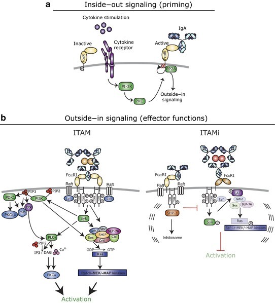 Fig 2. Model for the role of FcαRI in mucosal and systemic immunity. (a) Homeostasis: SIgA prevents bacterial invasion; serum IgA maintains anti-inflammatory tone via ITAMi. (b) Local translocation: dIgA opsonizes pathogens; neutrophils clear infection via ITAM signaling and LTB4 release. (c) Systemic translocation: serum IgA-opsonized bacteria are cleared by Kupffer cells. (Source: van der Steen et al., Mucosal Immunology 2011)
FcαR in Mucosal Immunity
Mucosal immunity represents the largest component of the human immune system, and IgA is its predominant antibody class. While secretory IgA (SIgA) provides frontline defense through immune exclusion, the Fcα receptor system operates as a critical second and third line of defense when pathogens breach epithelial barriers.
In homeostatic conditions, dimeric IgA (dIgA) produced by lamina propria plasma cells is transported across epithelial cells via the polymeric Ig receptor (pIgR) to become SIgA. SIgA effectively prevents microbial attachment and neutralizes toxins without triggering inflammation. However, when microorganisms penetrate the epithelial barrier, dIgA in the lamina propria can opsonize these pathogens. Recruited neutrophils expressing FcαRI recognize the IgA-coated microbes and initiate clearance through phagocytosis and the release of leukotriene B4 (LTB4), establishing a self-controlled positive feedback loop until the infection is resolved.
Recent studies have revealed that CD89 is upregulated on subsets of mucosal NK cells, particularly in cord blood and mucosal tissues. These CD89+ NK cells exhibit a unique signaling profile, utilizing CD3ζ, Syk, and ZAP70, but also express high levels of inhibitory receptors such as KLRG1. This suggests that CD89 on mucosal NK cells likely serves as a regulatory receptor, modulating immune responses depending on IgA subclass and the presence of other antibody isotypes. Notably, combination IgG and IgA2 antibody-dependent cellular cytotoxicity (ADCC) generates enhanced NK cell responses compared to IgG alone, indicating complex cross-isotype regulation at mucosal surfaces.
Three Lines of Mucosal Defense:
1. First Line (Immune Exclusion): SIgA prevents microbial attachment and neutralizes pathogens in the lumen without inflammation.
2. Second Line (Local Clearance): When barriers are breached, dIgA opsonizes pathogens; neutrophils via FcαRI mediate phagocytosis and LTB4-driven recruitment.
3. Third Line (Systemic Defense): Serum IgA-opsonized bacteria entering portal circulation are cleared by FcαRI+ Kupffer cells in the liver.
FcαR in Inflammation and Autoimmune Research
The IgA-FcαRI axis plays a paradoxical role in inflammation and autoimmunity, capable of both protecting against and promoting tissue damage. Aberrant IgA immune complex formation or impaired clearance can lead to excessive FcαRI-mediated neutrophil activation, resulting in severe tissue injury across multiple disease contexts.
IgA nephropathy (IgAN) exemplifies the pathological potential of dysregulated IgA-FcαRI interactions. Elevated serum IgA levels, IgA autoantibodies, and circulating immune complexes are hallmark features. The sCD89-TfR1 axis contributes to mesangial IgA deposition, and therapeutic strategies targeting this pathway—including mTOR inhibitors like everolimus—show promise in correcting receptor dysfunctions and slowing disease progression.
Other autoimmune and inflammatory conditions associated with altered IgA-CD89 biology include rheumatoid arthritis, IgA vasculitis, dermatitis herpetiformis, celiac disease, inflammatory bowel disease, Sjögren's syndrome, and ankylosing spondylitis. In these diseases, excessive IgA immune complexes may trigger disproportionate myeloid cell activation, releasing pro-inflammatory cytokines (TNF-α, IL-1β, IL-6) and reactive oxygen species that drive tissue destruction.
Conversely, the anti-inflammatory ITAMi signaling induced by monomeric IgA presents therapeutic opportunities. Inducing FcαRI-mediated inhibitory signals could potentially reduce allergic inflammation or autoimmune pathology. Additionally, blocking FcαRI with monoclonal antibodies or peptides may resolve IgA-induced tissue damage in hyper-inflammatory states.
Recombinant CD89/FCAR Protein Applications
Recombinant CD89/FCAR proteins are invaluable tools for dissecting IgA-mediated immune mechanisms and developing novel therapeutic strategies. These proteins enable researchers to study ligand-binding interactions, signaling pathways, and cellular activation in controlled experimental settings.
Research Applications
- In vitro IgA-mediated activation studies: Recombinant CD89 can be used as a soluble factor or coating matrix protein to investigate inflammation pathway regulation and FcαRI-mediated cell activation.
- Protein-protein interaction mapping: Structural and binding studies using recombinant FcαRI domains to map IgA interaction sites and identify competing ligands.
- Biomarker development: Soluble CD89 (sCD89) serves as a potential biomarker for monitoring IgA-mediated inflammatory pathway status in diseases such as IgA nephropathy.
- Immunogen for antibody production: Recombinant CD89 protein can be used to generate highly specific monoclonal and polyclonal antibodies for research and diagnostic applications.
- Bispecific antibody engineering: CD89-targeting bispecific constructs can recruit neutrophils and macrophages for potent antibody-dependent cellular cytotoxicity against tumor cells or pathogens.
Therapeutic Potential
The unique biology of the IgA receptor opens several therapeutic avenues. IgA monoclonal antibodies directed against tumor antigens may leverage the superior neutrophil-killing capacity of IgA compared to IgG. Furthermore, FcαRI-targeted bispecific antibodies can redirect myeloid effector cells to eliminate malignant cells. On the other hand, FcαRI blockade strategies may benefit patients with IgA-driven autoimmune diseases by preventing immune complex-mediated tissue damage.
| Application Area | Recombinant CD89 Format | Purpose |
|---|---|---|
| Signal transduction research | Full-length extracellular domain (ECD) | Study IgA binding kinetics and receptor dimerization |
| Drug screening | Soluble FcαRI-Fc fusion protein | Identify small molecules or peptides that modulate IgA binding |
| Cell-based assays | His-tagged or Fc-tagged CD89 | Coat plates for neutrophil/monocyte activation studies |
| Structural biology | ECD with defined glycosylation | Crystallography and cryo-EM studies of IgA-FcαRI complexes |
| Diagnostic development | sCD89 ELISA standards | Quantify soluble CD89 in patient serum as disease biomarker |
Creative BioMart Fcα Receptor Solutions
Creative BioMart offers a comprehensive portfolio of Fc receptor products to support your IgA-mediated immunity research. Our CD89/FCAR protein catalog includes high-purity recombinant proteins produced in multiple expression systems, ensuring native-like glycosylation and proper folding for reliable experimental results.
Explore Our Fcα Receptor Product Line
Learn More
View All Fc Receptors
Learn More
Our Fc alpha receptor solutions include:
- Recombinant Human CD89/FCAR Proteins: Available with His-tag, Fc-tag, or tag-free formats; produced in HEK293, CHO, or insect cells for optimal activity.
- Anti-CD89 Antibodies: Monoclonal and polyclonal antibodies validated for flow cytometry, Western blot, IHC, and functional blocking assays.
- Fc Receptor Multiplex Assays: High-throughput screening tools for profiling Fc receptor binding across antibody isotypes.
- Custom Protein Services: Tailored protein engineering, including mutant variants, domain truncations, and species orthologs (mouse, rat, cynomolgus).
Whether you are investigating mucosal immunity, developing IgA-based therapeutics, or studying autoimmune disease mechanisms, Creative BioMart provides the reliable reagents and expert support you need to advance your research.
Resource
-
What Are Fc Receptors? The Immune System's Antibody Response Switch
-
FcRn: The Secret to Long-Lasting Antibodies!
-
ADCC vs ADCP: How Fcγ Receptors Drive Cancer Immunity
-
CD64: The Tiny Receptor with Big Implications!
-
Fc Receptor Polymorphisms: Why Antibody Responses Differ Between Patients
References
- Herr AB, Ballister ER, Bjorkman PJ. Insights into IgA-mediated immune responses from the crystal structures of human FcαRI and its complex with IgA1-Fc. Nature. 2003;423(6938):614-620. doi:10.1038/nature01685
- van der Steen L, Tuk CW, Bakema JE, et al. The human immunoglobulin A Fc receptor FcαRI: a multifaceted regulator of mucosal immunity. Mucosal Immunol. 2011;4(6):612-624. doi:10.1038/mi.2011.36
- Kroll KW, Hueber B, Balachandran H, et al. FcαRI (CD89) is upregulated on subsets of mucosal and circulating NK cells and regulates IgA-class specific signaling and functions. Mucosal Immunol. 2024;17(4):692-699. doi:10.1016/j.mucimm.2024.04.003
- Pasquier B, Launay P, Kanamaru Y, et al. Identification of FcαRI as an inhibitory receptor that controls inflammation: dual role of FcRγ ITAM. Immunity. 2005;22(1):31-42. doi:10.1016/j.immuni.2004.11.017
- Ben Mkaddem S, Benhamou M, Monteiro RC. Anti-inflammatory role of the IgA Fc receptor (CD89): from autoimmunity to therapeutic perspectives. Autoimmun Rev. 2013;12(6):666-669. doi:10.1016/j.autrev.2012.10.011
- Brandsma AM, Bondza S, Evers M, et al. Potent Fc Receptor Signaling by IgA Leads to Superior Killing of Cancer Cells by Neutrophils Compared to IgG. Front Immunol. 2019;10:704. doi:10.3389/fimmu.2019.00704
- van Egmond M, van Garderen E, van Spriel AB, et al. FcαRI-positive liver Kupffer cells: reappraisal of the function of immunoglobulin A in immunity. Nat Med. 2000;6(6):680-685. doi:10.1038/76261
- Chen A, Hsu CY, Chen YC, et al. IgA nephropathy: clearance kinetics of IgA-containing immune complexes. Semin Immunopathol. 2018;40(6):539-543. doi:10.1007/s00281-018-0708-7
- Bakema JE, van Egmond M. IgA and FcαRI: pathological roles and therapeutic opportunities. Front Immunol. 2019;10:553. doi:10.3389/fimmu.2019.00553
- Aleyd E, van Hout MW, Ganzevles SH, et al. IgA enhances NETosis and release of neutrophil extracellular traps by polymorphonuclear cells via Fcα receptor I. J Immunol. 2014;192(5):2374-2383. doi:10.4049/jimmunol.130.0589
- Macpherson AJ, Geuking MB, McCoy KD. Homeland Security: IgA immunity at the frontiers of the body. Trends Immunol. 2012;33(4):160-167. doi:10.1016/j.it.2012.01.006
- Monteiro RC, Van De Winkel JG. IgA Fc receptors. Annu Rev Immunol. 2003;21:177-204. doi:10.1146/annurev.immunol.21.120601.141011

Contact us or send an email at for project quotations and more detailed information.
Quick Links
-

Papers’ PMID to Obtain Coupon
Submit Now -

Refer Friends & New Lab Start-up Promotions