

Fcε Receptor Proteins for Allergy and IgE Research
A comprehensive resource on FcεRI, FcεRII/CD23, and recombinant proteins for understanding IgE-mediated allergic inflammation, mast cell activation, and therapeutic development in asthma and hypersensitivity disorders.
Table of Contents
Overview of Fcε Receptors
Fc Receptors (FcRs) are a family of cell surface glycoproteins that bind the Fc region of immunoglobulins, bridging the adaptive and innate immune systems. Among these, Fc epsilon receptors (FcεRs) are uniquely dedicated to binding immunoglobulin E (IgE) and play a central role in type I hypersensitivity reactions, including allergic asthma, rhinitis, urticaria, and anaphylaxis.
Two principal classes of Fcε receptors have been characterized: FcεRI (the high-affinity IgE receptor) and FcεRII (also known as CD23, the low-affinity IgE receptor). While both receptors recognize the Fc portion of IgE, they differ dramatically in binding affinity, cellular distribution, structural architecture, and biological function. Understanding these differences is fundamental to dissecting the mechanisms of IgE-mediated allergic diseases and developing targeted therapeutics.
Key Facts About Fcε Receptors
- FcεRI binds IgE with extraordinarily high affinity (KD ≈ 10⁻¹⁰ M), essentially irreversible under physiological conditions.
- FcεRII/CD23 binds IgE with low affinity (KD ≈ 10⁻⁸ M) and exists in both membrane-bound and soluble forms.
- FcεRI expression is upregulated by IgE itself, creating a positive feedback loop that amplifies allergic sensitization.
- Genetic polymorphisms in FCER1A, FCER1G, and FCER2 have been associated with atopy, asthma severity, and IgE levels.
FcεRI vs FcεRII/CD23
The distinction between FcεRI and FcεRII extends beyond simple affinity differences. These receptors operate in distinct cellular contexts and mediate different aspects of the allergic response. The following table summarizes their key characteristics:
| Feature | FcεRI (High-Affinity) | FcεRII/CD23 (Low-Affinity) |
|---|---|---|
| Gene Symbols | FCER1A, FCER1B, FCER1G | FCER2 |
| Binding Affinity (KD) | ~10⁻¹⁰ M (high affinity) | ~10⁻⁸ M (low affinity) |
| Structure | Tetrameric (αβγ₂) or trimeric (αγ₂) | Type II transmembrane C-type lectin; exists as trimer |
| Primary Cells | Mast cells, basophils, DCs, Langerhans cells, monocytes | B cells, macrophages, eosinophils, follicular DCs, epithelial cells |
| IgE Binding Site | Cε3-Cε4 domains of IgE Fc | Cε3 domain of IgE Fc |
| Signaling | ITAM-mediated (β and γ chains); triggers degranulation | No ITAM; regulates IgE synthesis, antigen presentation |
| Biological Role | Immediate hypersensitivity; mast cell/basophil activation | IgE homeostasis; B cell differentiation; non-inflammatory antigen transport |
| Therapeutic Relevance | Primary target for anti-IgE therapies (e.g., omalizumab, ligelizumab) | Potential target for IgE regulation; biomarker for asthma |
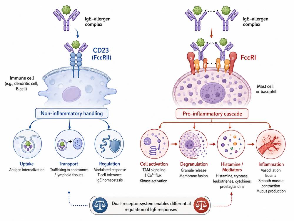 Fig 1. CD23 (FcεRII) provides a non-inflammatory pathway for IgE-allergen complexes, in contrast to the pro-inflammatory FcεRI-mediated cascade. This dual-receptor system allows differential regulation of IgE responses.
The FCER1A-encoded α chain of FcεRI contains two extracellular Ig-like domains that form the complete IgE-binding site. In contrast, FCER2-encoded CD23 is a type II membrane protein with a C-terminal C-type lectin domain responsible for IgE recognition. Notably, CD23 can trap IgE in a "closed" conformation that prevents its interaction with FcεRI, offering an endogenous regulatory mechanism that has inspired therapeutic strategies.
Fcε Receptors in Mast Cells and Basophils
Mast cells and basophils are the primary effector cells of IgE-mediated allergic reactions, and their function is critically dependent on Fc Receptors, particularly FcεRI. On these cells, FcεRI exists predominantly as a heterotetramer (αβγ₂), though a trimeric form (αγ₂) lacking the β chain is found on antigen-presenting cells such as dendritic cells and Langerhans cells.
FcεRI Structure and Assembly
The tetrameric FcεRI complex is assembled from:
- α chain (FCER1A): Contains the extracellular IgE-binding domain, a transmembrane region, and a short cytoplasmic tail. It is the only subunit that directly contacts IgE.
- β chain (FCER1B/MS4A2): A four-transmembrane protein that amplifies signaling and stabilizes the receptor complex.
- γ chains (FCER1G): A homodimer, each subunit contains an immunoreceptor tyrosine-based activation motif (ITAM) essential for signal transduction.
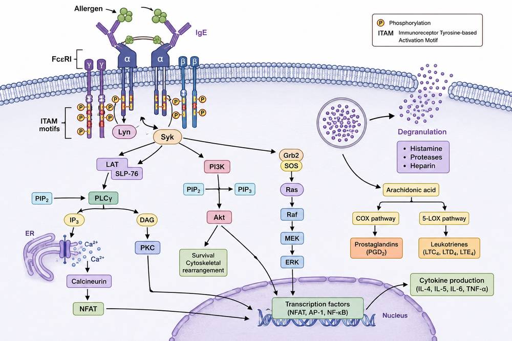 Fig 2. FcεRI signaling cascade in mast cells. Upon allergen-mediated crosslinking, Lyn and Syk kinases phosphorylate ITAM motifs, activating downstream pathways (PLCγ, PI3K, MAPK) that drive degranulation, eicosanoid synthesis, and cytokine transcription.
Signal Transduction Mechanism
In resting mast cells and basophils, FcεRI is constitutively expressed and loaded with monomeric IgE. The binding of multivalent allergen to adjacent IgE molecules induces receptor aggregation, which initiates a rapid signaling cascade:
- Lyn kinase associates with the β chain and phosphorylates ITAMs on both β and γ chains.
- Syk kinase is recruited to phosphorylated γ-chain ITAMs, becoming activated.
- Activated Syk phosphorylates LAT (Linker for Activation of T cells) and SLP-76, creating docking sites for downstream effectors.
- PLCγ activation generates IP₃ and DAG, triggering Ca²⁺ mobilization and PKC activation.
- Concurrently, the PI3K-Akt and Ras-MAPK pathways are activated, leading to NF-κB nuclear translocation.
This integrated signaling network culminates in three major outcomes: (1) immediate release of preformed granule mediators (histamine, proteases, heparin); (2) de novo synthesis of lipid mediators (leukotrienes, prostaglandins, PAF); and (3) transcription and secretion of cytokines and chemokines (IL-4, IL-5, IL-13, TNF-α).
Beyond Mast Cells and Basophils
Although mast cells and basophils express the highest levels of FcεRI, the receptor is also present—at 10- to 100-fold lower density—on dendritic cells, Langerhans cells, eosinophils, monocytes, and bronchial epithelial cells from asthmatic patients. On these cells, FcεRI functions primarily in antigen presentation and immune modulation rather than degranulation, highlighting the receptor's versatility in allergic inflammation.
IgE Binding and Allergic Inflammation
The interaction between IgE and Fc Receptors is the molecular cornerstone of allergic disease. IgE binds FcεRI with a dissociation constant of approximately 10⁻¹⁰ M, making this one of the strongest non-covalent protein-protein interactions known in immunology. This extraordinary affinity means that IgE essentially remains bound to FcεRI for the lifetime of the mast cell or basophil, effectively "arming" these cells for rapid allergen response.
The Allergic Cascade
The sequence of IgE-mediated allergic inflammation can be summarized as follows:
Phase 1: Sensitization
Antigen-presenting cells process allergens and present them to Th2 cells, which drive B cell class switching to IgE production. Secreted IgE circulates and binds to FcεRI on mast cells and basophils.
Phase 2: Effector Response (Early Phase)
Upon re-exposure, multivalent allergen crosslinks FcεRI-bound IgE, triggering degranulation within seconds to minutes. Histamine causes vasodilation, vascular permeability, and smooth muscle contraction. Lipid mediators amplify and sustain the response.
Phase 3: Late-Phase Reaction
Hours after initial exposure, newly synthesized cytokines (IL-4, IL-5, IL-13, TNF-α) and chemokines recruit eosinophils, neutrophils, and Th2 cells, establishing chronic allergic inflammation.
Glycosylation Modulates IgE-FcεRI Interaction
Recent research has revealed that IgE glycosylation—specifically sialylation—significantly modulates its interaction with FcεRIα. Higher sialylation of IgE reduces binding to FcεRI and attenuates mast cell and basophil activation, while desialylated IgE shows enhanced receptor interaction and cellular activation. This finding opens new avenues for understanding disease heterogeneity and developing glycoengineered therapeutic approaches.
Soluble FcεRIα (sFcεRIα)
A soluble form of the FcεRI α chain can be detected in human serum. sFcεRIα retains IgE-binding capacity and may function as a decoy receptor, sequestering free IgE and preventing its binding to cellular FcεRI. Levels of sFcεRIα correlate with total serum IgE and may serve as a biomarker for allergic disease activity. Notably, some studies suggest elevated sFcεRIα in tolerant subjects compared to allergic individuals, hinting at a potential protective role.
FcεR in Asthma and Hypersensitivity Research
Allergic asthma is a chronic inflammatory airway disease driven by type 2 immune responses, with the IgE-FcεRI axis serving as a central pathogenic mechanism. In asthmatic patients, FcεRI expression is upregulated not only on mast cells and basophils but also on bronchial epithelial cells, dendritic cells, and monocytes, creating a pro-inflammatory environment throughout the airway mucosa.
FcεRI as a Therapeutic Target in Asthma
Targeting the IgE-FcεRI protein-protein interaction (PPI) represents one of the most validated strategies in allergic disease therapy:
- Omalizumab (anti-IgE monoclonal antibody) binds free IgE, preventing its interaction with FcεRI and reducing receptor expression on effector cells. It is approved for severe allergic asthma and chronic spontaneous urticaria.
- Ligelizumab (QGE031) is a next-generation anti-IgE antibody with higher affinity for IgE, currently under investigation for food allergy and asthma.
- ECRIs (Allergic Effector Cell Response Inhibitors) represent a novel class of therapeutics that not only neutralize free IgE but also remove cell-bound IgE and downregulate FcεRI expression, offering a comprehensive "disarming" strategy.
FCER2/CD23 in Asthma Genetics
Genetic studies have identified single nucleotide polymorphisms (SNPs) in FCER2 that are associated with asthma severity, IgE levels, and response to corticosteroid therapy. The FCER2 T2206C polymorphism affects CD23 stability and IgE binding, influencing disease phenotype and therapeutic outcomes. These findings underscore the clinical relevance of FcεRII as both a biomarker and potential therapeutic target.
Type I Hypersensitivity and Beyond
Beyond asthma, FcεRI signaling drives type I hypersensitivity reactions across multiple organ systems: allergic rhinitis (nasal mast cells), atopic dermatitis (skin mast cells), food allergy (intestinal mast cells), and systemic anaphylaxis (circulating basophils and tissue mast cells). The central role of FcεRI in these diverse pathologies makes it an attractive target for broad-spectrum anti-allergic therapeutics.
Recombinant FCER1A, FCER1G, and FCER2 Proteins
High-quality recombinant FCER1A, FCER1G, and FCER2 proteins are essential tools for allergy and immunology research. Creative BioMart offers a comprehensive portfolio of recombinant Fcε receptor proteins produced in mammalian (HEK293) and bacterial (E. coli) expression systems, with various tags and modifications to suit diverse experimental needs.
🔳 Protein Selection Guide
- For IgE binding studies: Use biotinylated or His-tagged FCER1A extracellular domain proteins.
- For signaling research: Combine FCER1A with FCER1G to reconstitute functional receptor complexes.
- For CD23/FcεRII studies: Use FCER2 proteins to investigate low-affinity IgE interactions and B cell regulation.
- For antibody discovery: Membrane protein formats (MP series) preserve native conformation for therapeutic antibody screening.
Applications in IgE Binding Assays
Recombinant Fcε receptor proteins are indispensable reagents for a wide range of IgE-focused research applications. Their defined composition, high purity, and batch-to-batch consistency enable quantitative and reproducible assays that are critical for both basic research and drug discovery.
1. Surface Plasmon Resonance (SPR) and Bio-Layer Interferometry (BLI)
Biotinylated FCER1A and FCER2 proteins can be immobilized on sensor chips to measure real-time binding kinetics of IgE antibodies, allergen-IgE complexes, or therapeutic candidates. These assays provide accurate determination of association rate constants (kon), dissociation rate constants (koff), and equilibrium dissociation constants (KD), which are essential for characterizing drug candidates targeting the IgE-FcεRI axis.
2. ELISA and Competitive Binding Assays
Recombinant Fcε receptor proteins serve as capture antigens in ELISA formats to detect and quantify IgE levels in serum or culture supernatants. In competitive formats, soluble receptor proteins can be used to assess the inhibitory potency of small molecules, peptides, or antibodies designed to disrupt IgE-FcεRI interactions.
3. Flow Cytometry and Cellular Assays
Fluorescently labeled FCER2 proteins (e.g., Alexa Fluor 488 conjugates) enable flow cytometric analysis of IgE-expressing B cells or CD23-expressing cell populations. Recombinant FCER1A proteins can be used to stain IgE-armed basophils and mast cells, providing a direct readout of receptor occupancy.
4. Protein Arrays and High-Throughput Screening
GST-tagged FCER1G and His-tagged FCER1A proteins are compatible with protein microarray platforms, enabling high-throughput screening of compound libraries for inhibitors of IgE-receptor binding. These platforms accelerate the discovery of small-molecule therapeutics for allergic diseases.
5. Structural Biology
High-purity recombinant Fcε receptor proteins are essential for crystallization and cryo-EM studies aimed at resolving the atomic structures of IgE-receptor complexes. Such structural insights have directly enabled the rational design of therapeutic antibodies (e.g., omalizumab, ligelizumab) and are guiding the development of next-generation inhibitors.
Creative BioMart Fcε Receptor Products
Creative BioMart is a leading supplier of recombinant Fc Receptor proteins for allergy, asthma, and immunology research. Our Fcε receptor product line includes:
- Recombinant Proteins: Full-length and extracellular domain proteins for FCER1A, FCER1G, and FCER2 from human, mouse, rat, and cynomolgus sources.
- Membrane Protein Formats: Native-like membrane proteins produced via in vitro wheat germ expression for antibody discovery and structural studies.
- Tagged Variants: His, GST, Fc, Avi, and biotinylated formats for flexible assay design.
- Custom Services: Protein engineering, stable cell line development, and assay development services tailored to your IgE/allergy research program.
Why Choose Creative BioMart Fcε Receptor Proteins?
- High Purity: >85% purity by SDS-PAGE; endotoxin levels <1 EU/µg.
- Verified Activity: Functional validation by IgE binding ELISA and SPR.
- Multiple Species: Human, mouse, rat, and non-human primate options for translational research.
- Flexible Formats: Soluble, membrane-bound, and conjugated proteins to match your assay requirements.
- Custom Production: Bulk quantities and custom tags available upon request.
Browse All Fc Receptor Products
Learn More
Resource
-
What Are Fc Receptors? The Immune System's Antibody Response Switch
-
FcRn: The Secret to Long-Lasting Antibodies!
-
ADCC vs ADCP: How Fcγ Receptors Drive Cancer Immunity
-
CD64: The Tiny Receptor with Big Implications!
-
Fc Receptor Polymorphisms: Why Antibody Responses Differ Between Patients
References
- Kinet JP. The high-affinity IgE receptor (FcεRI): from physiology to pathology. Annu Rev Immunol. 1999;17:931-972.
- Gould HJ, Sutton BJ. IgE in allergy and asthma today. Nat Rev Immunol. 2008;8(3):205-217.
- Blank U, Rivera J. The ins and outs of IgE-dependent mast-cell exocytosis. Trends Immunol. 2004;25(5):266-273.
- Platzer B, Ruiter F, van der Molen R, et al. Soluble FcεRIα generates negative feedback activation of human basophils. J Allergy Clin Immunol. 2011;128(2):429-435.
- Dehlink E, Platzer B, Baker AH, et al. Solable FcεRIα: an endogenous immunoregulator in allergic disease? Clin Exp Allergy. 2011;41(7):943-951.
- Holgate ST, Polosa R. The mechanisms, diagnosis, and management of severe asthma in adults. Lancet. 2006;368(9537):780-793.
- Beck LA, Marcotte GV, MacGlashan D, et al. Omalizumab-induced reductions in mast cell FcεRI expression and function. J Allergy Clin Immunol. 2004;114(3):527-530.
- Davies AM, Allan EG, Keeble AH, et al. Allosteric mechanism of action of the therapeutic anti-IgE antibody omalizumab. J Biol Chem. 2017;292(24):9975-9987.
- Liu YJ, Wang HY, Wang R, et al. IgE-FcεRI protein-protein interaction as a therapeutic target against allergic asthma: An updated review. Int Immunopharmacol. 2025;139:113610.
- Harris G. Validating the trifunctional mechanism of action of Effector Cell Response Inhibitors (ECRIs). Presented at ACAAI 2025, Orlando, FL.
- Henningsson F, Yamamoto K, Saarinen JV, et al. Sialylation of IgE reduces FcεRIα interaction and mast cell and basophil activation in vitro and increases IgE half-life in vivo. Allergy. 2023;78(2):456-469.
- Greer AM, Wu N, Putnam AL, et al. Serum IgE clearance is facilitated by human FcεRI internalization. J Clin Invest. 2014;124(3):1187-1198.
- Tantisira KG, Lasky-Su J, Harada M, et al. Genomewide association between GLCCI1 and response to glucocorticoid therapy in asthma. N Engl J Med. 2011;365(13):1173-1183.
- Takai T. Fc receptors and their role in immune regulation and autoimmunity. J Clin Immunol. 2005;25(1):1-18.

Contact us or send an email at for project quotations and more detailed information.
Quick Links
-

Papers’ PMID to Obtain Coupon
Submit Now -

Refer Friends & New Lab Start-up Promotions