

CD16a / FcγRIIIa Proteins for ADCC Assay Development
High-quality recombinant CD16a proteins engineered for antibody-dependent cellular cytotoxicity (ADCC) assays, monoclonal antibody screening, and Fc engineering applications.
Table of Contents
- Why CD16a Matters in ADCC
- CD16a Expression on NK Cells
- CD16a–IgG Fc Binding Mechanism
- CD16a Variants and Antibody Binding Differences
- Applications in Monoclonal Antibody Screening
- Applications in Fc Engineering
- Recombinant CD16a Protein Formats
- How to Choose CD16a Proteins for Binding Assays
- Creative BioMart CD16/CD16a Product Options
Why CD16a Matters in ADCC
Antibody-dependent cellular cytotoxicity (ADCC) is a critical effector mechanism by which therapeutic monoclonal antibodies (mAbs) eliminate target cells, particularly in oncology and infectious disease applications. At the center of this process is CD16a (also known as FcγRIIIa), the low-affinity Fc receptor expressed predominantly on natural killer (NK) cells.
CD16a serves as the primary bridge between the Fc region of IgG antibodies bound to target cells and the cytotoxic machinery of NK cells. When an antibody's Fab regions recognize antigens on a tumor cell or pathogen-infected cell, the Fc domain engages CD16a on adjacent NK cells. This cross-linking triggers a signaling cascade through the associated FcεRI-γ chain, leading to the release of perforin, granzymes, and pro-inflammatory cytokines that ultimately lyse the target cell.
Key Insight: The affinity of CD16a for the Fc region of therapeutic antibodies directly correlates with ADCC potency. A positive correlation between CD16a affinity and ADCC activity has been demonstrated across multiple studies, making CD16a binding assays an essential early readout in antibody drug development.
Unlike other Fcγ receptors such as FcγRI (CD64) or FcγRII (CD32), CD16a is uniquely positioned as the dominant activating receptor on NK cells for mediating ADCC. Its medium affinity for IgG allows it to bind immune complexes efficiently while avoiding saturation by circulating monomeric IgG, making it the ideal sensor for antibody-opsonized target cells.
CD16a Expression on NK Cells
CD16a is the most abundant activating Fc receptor on human NK cells, typically expressed on >90% of circulating CD56dim NK cells—the subset responsible for the majority of ADCC activity. CD16a surface density on NK cells ranges from approximately 50,000 to over 100,000 receptors per cell, depending on the individual's FCGR3A genotype.
CD16a expression is not static; it is dynamically regulated by activation state. Upon engagement with antibody-opsonized targets, CD16a is rapidly downregulated from the NK cell surface through ectodomain shedding (mediated by ADAM17). This shedding is functionally important—it facilitates NK cell detachment from the target, enabling the effector cell to engage multiple targets sequentially in a process known as "serial killing."
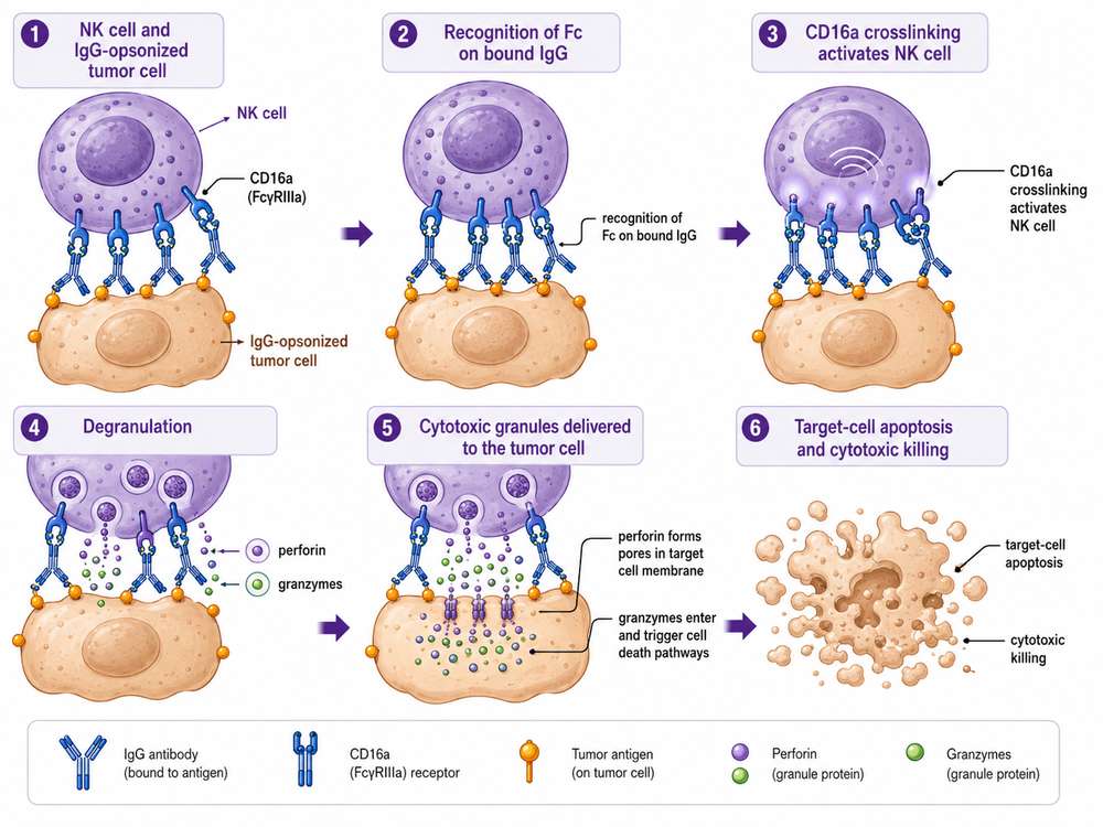 Fig 1. CD16a (FcγRIIIa) on NK cells recognizes antibody-opsonized tumor cells and triggers ADCC through granule release and direct cytotoxicity.
Beyond NK cells, CD16a is also expressed on activated monocytes, macrophages, and a subset of γδ T cells, though NK cells remain the primary mediators of ADCC in therapeutic antibody contexts. The restricted expression pattern of CD16a makes it an attractive target for both assay development and therapeutic engineering.
CD16a–IgG Fc Binding Mechanism
The interaction between CD16a and the Fc region of IgG antibodies is a well-characterized protein-protein interaction that serves as the molecular basis for ADCC. The binding interface involves the membrane-proximal IgG-binding domain of CD16a and the CH2-CH3 hinge region of the IgG Fc fragment.
Key structural features of the CD16a–IgG Fc interaction include:
- CH2 domain contact: The lower hinge and CH2 domain of IgG Fc form the primary binding surface for CD16a
- Glycan-mediated interaction: The N-linked glycan at Asn297 of the IgG Fc is essential for optimal CD16a binding. The absence of core fucose (afucosylation) dramatically increases affinity
- Two-site binding model: CD16a engages both CH2 domains of the Fc simultaneously, with each heavy chain contributing to the binding interface
- Key CD16a residues: Tyr132, Trp90, and Trp113 in the CD16a extracellular domain are critical contact residues that influence binding affinity
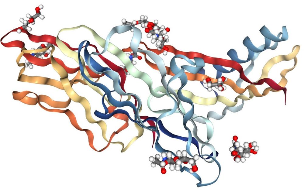 Fig 2. Crystal structure of CD16a (FcγRIIIa) showing the extracellular domain architecture critical for IgG Fc engagement.
The binding affinity of CD16a for IgG1 Fc is typically in the low micromolar range (KD ~1–10 μM for the F176 variant), which is characteristic of a medium-affinity receptor. This affinity is sufficient to support stable immune synapse formation when multiple CD16a molecules engage antibody-opsonized targets, while still allowing for dynamic regulation and serial killing.
CD16a Variants and Antibody Binding Differences
The FCGR3A gene encoding CD16a contains several clinically relevant polymorphisms that significantly impact antibody binding affinity and ADCC activity. The most extensively studied variant is the 158V/F polymorphism (also referred to as the 176V/F polymorphism depending on the numbering system used).
FcγRIIIa-158V/F Polymorphism
A single nucleotide polymorphism (SNP) at position 559 in the FCGR3A gene results in an amino acid substitution at position 158: valine (V) or phenylalanine (F). This residue lies within the IgG-binding domain and directly influences the affinity for IgG Fc:
| Genotype | CD16a Allotype | Relative IgG1 Affinity | ADCC Activity | Clinical Relevance |
|---|---|---|---|---|
| V/V (homozygous) | High-affinity V158 | Highest (~4× higher than F/F) | Strongest ADCC | Improved rituximab response in lymphoma |
| V/F (heterozygous) | Intermediate | Intermediate | Moderate ADCC | Intermediate clinical outcomes |
| F/F (homozygous) | Low-affinity F158 | Lowest | Weakest ADCC | Reduced rituximab efficacy |
Studies have shown that NK cells from V/V homozygous donors exhibit approximately 4-fold higher affinity for IgG1 compared to F/F homozygotes, with corresponding differences in ADCC-mediated killing. In clinical settings, patients with follicular lymphoma carrying the V/V genotype demonstrated improved response rates and event-free survival following rituximab treatment compared to F/F carriers.
Assay Implication: When developing ADCC assays or screening therapeutic antibodies, it is critical to evaluate binding to both CD16a-158V and CD16a-158F variants. Antibodies engineered to enhance CD16a binding should ideally show improved affinity for the low-affinity F/F variant to broaden therapeutic efficacy across patient genotypes.
Additional Polymorphisms
Beyond the 158V/F variant, the 48L/R/H polymorphism in the membrane-distal domain and the 176V/F polymorphism (equivalent to 158V/F in alternate numbering) also influence CD16a function. The L48H/R variant has been associated with enhanced serial killing capacity by promoting more efficient NK cell detachment from target cells.
Applications in Monoclonal Antibody Screening
Recombinant CD16a protein is an indispensable reagent in the early-stage screening and characterization of therapeutic monoclonal antibodies. By measuring the binding affinity of candidate mAbs to CD16a, developers can rank-order lead molecules based on predicted ADCC potency before proceeding to costly cell-based assays.
Common assay formats using recombinant CD16a protein for mAb screening include:
| Assay Format | Readout | Advantages | Typical Application |
|---|---|---|---|
| SPR (Biacore) | Kon, Koff, KD | Label-free, real-time kinetics | Detailed affinity characterization |
| BLI (Octet) | Kon, Koff, KD | High throughput, minimal sample | Screening large antibody panels |
| ELISA | EC50, binding curve | Simple, cost-effective | Primary screening, lot release |
| AlphaLISA/TR-FRET | Affinity ranking | Homogeneous, high throughput | Large-scale library screening |
These binding assays serve as critical quality control checkpoints during the CMC (Chemistry, Manufacturing, and Controls) stage of antibody drug development. Batch-to-batch consistency of CD16a protein reagents ensures reproducible affinity measurements, which is essential for regulatory submissions and product release.
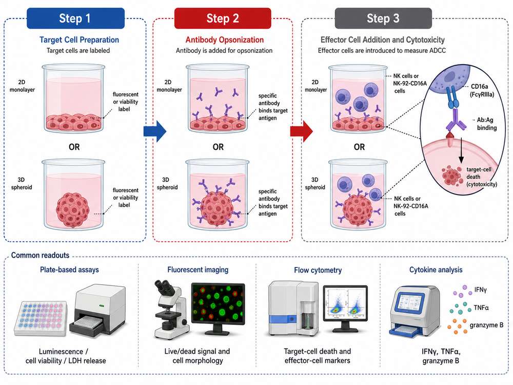 Fig 3. Standard ADCC assay workflow: target cells are labeled, antibody is added for opsonization, and effector cells (NK cells or NK-92-CD16A cells) are introduced to measure cytotoxicity.
Applications in Fc Engineering
Fc engineering represents one of the most impactful strategies for enhancing the therapeutic efficacy of monoclonal antibodies. By modifying the Fc region to increase affinity for CD16a, developers can amplify ADCC activity without altering the antibody's antigen-binding specificity.
Glycoengineering: Afucosylation
The most clinically validated Fc engineering approach is afucosylation—the removal of core fucose from the N-linked glycan at Asn297. Afucosylated antibodies bind CD16a with dramatically increased affinity (up to 50-fold higher in some cases) and elicit significantly enhanced ADCC.
Key findings from afucosylation studies:
- Afucosylated anti-CD20 antibodies (e.g., obinutuzumab) demonstrate superior B-cell depletion compared to fucosylated counterparts
- The EC50 for CD107a externalization (a marker of NK cell degranulation) differs by approximately 20-fold between wild-type and glycoengineered antibodies
- Afucosylation enhances serial killing capacity by promoting more rapid NK cell detachment from target cells
- Enhanced CD16a binding can partially compensate for the reduced affinity associated with the low-affinity F/F patient genotype
Fc Amino Acid Mutagenesis
Site-directed mutagenesis of specific residues in the Fc CH2 domain (e.g., S239D/I332E, known as the "SDIE" mutation) can also enhance CD16a binding. These mutations create additional favorable contacts with the CD16a binding interface without affecting the overall IgG structure or serum half-life.
Engineering Strategy: When optimizing antibodies for ADCC, developers typically screen both glycoengineered (afucosylated) and mutagenized Fc variants using recombinant CD16a binding assays. The lead candidate is selected based on the optimal balance of CD16a affinity, FcγRIIb (inhibitory receptor) binding, and serum stability.
Recombinant CD16a Protein Formats
Recombinant CD16a protein is produced as the soluble extracellular domain (typically amino acids Gly17–Gln208) to preserve the native IgG-binding interface while eliminating the transmembrane and cytoplasmic regions. Multiple formats are available to suit different assay requirements:
| Format | Tag | Key Features | Best For |
|---|---|---|---|
| His-Tag | C-terminal 6×His or 8×His | Easy immobilization on Ni-NTA surfaces; simple purification | SPR, BLI, ELISA |
| Biotinylated (Avi-Tag) | His-Avi dual tag | Site-specific biotinylation; uniform orientation on streptavidin surfaces | High-precision SPR/BLI, bead-based assays |
| Fc Fusion | Human IgG1 Fc | Increased stability and half-life; dimeric structure | Cell-based assays, prolonged studies |
| HSA Fusion | Human Serum Albumin | Enhanced solubility and stability; reduced aggregation | Long-term storage, challenging assays |
| Fluorophore-Conjugated | R-PE, APC, FITC, etc. | Direct detection in flow cytometry and imaging | Flow cytometry binding assays |
Explore Our CD16a Proteins
Learn More
Expression Systems
CD16a is a heavily glycosylated protein, with N-linked glycans contributing to its apparent molecular weight of approximately 45–60 kDa (compared to a predicted ~24 kDa unglycosylated mass). Proper glycosylation is essential for native folding and IgG-binding activity. For this reason, HEK293 cells are the preferred expression system for recombinant CD16a production, as they provide mammalian-type glycosylation patterns that closely mimic native NK cell-derived CD16a.
Quality specifications for research-grade CD16a protein typically include:
- Purity: ≥95% by SDS-PAGE; ≥90% monomer by SEC-HPLC or SEC-MALS
- Endotoxin: <1.0 EU/μg (preferably <0.1 EU/μg for sensitive assays)
- Activity validation: Confirmed binding to human IgG1 Fc or therapeutic antibodies (bevacizumab, rituximab, trastuzumab) by SPR or BLI
- Batch consistency: Lot-specific COA with binding affinity data
How to Choose CD16a Proteins for Binding Assays
Selecting the appropriate recombinant CD16a protein format and variant is critical for generating meaningful and reproducible ADCC assay data. Consider the following decision criteria:
1. Variant Selection (158V vs. 158F)
For comprehensive antibody characterization, both CD16a-158V (high-affinity) and CD16a-158F (low-affinity) variants should be tested. If screening for broad patient population efficacy, prioritize the F variant—antibodies that show enhanced binding to the low-affinity F allotype are more likely to deliver consistent clinical outcomes across genotypes.
2. Tag Selection
- His-Tag: Versatile and cost-effective. Suitable for most ELISA, SPR, and BLI applications where anti-His capture surfaces are used.
- Biotinylated (Avi-Tag): Ideal for applications requiring uniform protein orientation, such as SPR on streptavidin sensor chips or bead-based flow cytometry assays. Site-specific biotinylation minimizes batch-to-batch variability.
- Fc-Tag or HSA-Tag: Consider when increased protein stability or dimeric avidity is desired, though these formats may introduce avidity artifacts in kinetic measurements.
3. Assay Platform Compatibility
| Assay Platform | Recommended CD16a Format | Capture Strategy |
|---|---|---|
| SPR | His-Tag or Biotinylated | Anti-His chip or SA chip |
| BLI | His-Tag or Biotinylated | His1K or SA biosensor |
| ELISA | His-Tag (any format) | Direct coating or capture |
| Flow Cytometry | Biotinylated or Fluorophore-conjugated | Streptavidin beads or direct staining |
| Alphascreen/TR-FRET | Biotinylated | Streptavidin donor beads |
Quality Control Requirements
Always verify that the CD16a protein has been validated for binding activity against a reference IgG1 antibody (e.g., bevacizumab or rituximab) with reported affinity constants. This ensures the protein is correctly folded and functionally active. Purity should be confirmed by both SDS-PAGE and SEC-MALS/SEC-HPLC to rule out aggregate contamination, which can cause non-specific binding artifacts.
Creative BioMart CD16/CD16a Product Options
Creative BioMart offers a comprehensive portfolio of recombinant CD16a and Fc receptor proteins specifically designed for ADCC assay development, antibody screening, and Fc engineering research. All products are manufactured under stringent quality control with validated bioactivity data.
Featured CD16a Products
| Cat.# | Product name | Source (Host) | Species | Tag | Protein Length |
|---|---|---|---|---|---|
| CD16a-0235H | Active Recombinant Human CD16a protein, His-tagged, Biotinylated | HEK293 | Human | His | Met1-Gln208,F176V |
Customization Services
Beyond catalog products, Creative BioMart provides flexible custom protein production services for CD16a and other Fc receptors:
- Species variants: Human, mouse, rat, cynomolgus monkey, rhesus macaque, and other species
- Expression systems: HEK293, CHO, insect cells, yeast, E. coli, and cell-free systems
- Tag options: His, Fc, GST, Flag, Myc, SUMO, Strep, Avi (biotinylated), and tag-free
- Conjugations: Biotin, R-PE, APC, FITC, Cy3, Cy5, Cy7, Alexa Fluor dyes
- Special formulations: Carrier-free, GMP-grade, custom buffers, endotoxin removal
Need Custom CD16a Protein for Your ADCC Assay?
Our protein engineering team can produce CD16a variants, mutants, and species orthologs tailored to your specific assay requirements. From microgram to gram-scale production with full QC validation.
Quality Commitment: All Creative BioMart CD16a proteins are manufactured with batch-to-batch consistency, validated by SPR/BLI for IgG binding activity, and supplied with comprehensive Certificates of Analysis (COA) including purity, endotoxin, and bioactivity data.
Related Products & Services
Resource
-
What Are Fc Receptors? The Immune System's Antibody Response Switch
-
FcRn: The Secret to Long-Lasting Antibodies!
-
ADCC vs ADCP: How Fcγ Receptors Drive Cancer Immunity
-
CD64: The Tiny Receptor with Big Implications!
-
Fc Receptor Polymorphisms: Why Antibody Responses Differ Between Patients

Contact us or send an email at for project quotations and more detailed information.
Quick Links
-

Papers’ PMID to Obtain Coupon
Submit Now -

Refer Friends & New Lab Start-up Promotions