


Fibroblast Growth Factor (FGF)
Related Symbol Search List
- FGF1
- FGF10
- FGF11
- FGF12
- FGF13
- FGF14
- FGF15
- FGF16
- FGF17
- FGF18
- FGF19
- FGF2
- FGF20
- FGF21
- FGF5
- FGF6
- FGF7
- FGF9
- FGFBP1
- FGFBP2
- FGFBP3
Immunology Background
Available Resources for FGF Research
Creative BioMart offers a wide range of assistance to researchers studying FGF ligands, providing a variety of products and a wealth of resources. These valuable assets serve as important references for researchers to help them better understand the critical functions of FGF ligands in a variety of physiological processes and diseases.
- Our product line includes recombinant proteins, magnetic beads pre-coupled to proteins, cell and tissue lysates, and more.
- Our resources related to FGF ligands include involved pathways, protein functions, interacting proteins, related articles, and research areas.
Our Featured Products
| Cat.# | Product name | Species | Source (Host) | Tag |
|---|---|---|---|---|
| FGF1-12861H | Recombinant Human FGF1, GST-tagged | Human | E.coli | GST |
| FGF2-001H | Recombinant Human FGF2 Protein, His-tagged | Human | E.coli | His |
| FGF6-12872H | Recombinant Human FGF6, GST-tagged | Human | E.coli | GST |
| FGF7-1900C | Recombinant Chicken FGF7 Protein, His tagged | Chicken | E.coli | N-His |
| FGF12-12862H | Recombinant Human FGF12, GST-tagged | Human | E.coli | GST |
| FGF13-12863H | Recombinant Human FGF13, GST-tagged | Human | E.coli | GST |
| FGF14-2327R | Recombinant Rat FGF14 Protein | Rat | Mammalian Cell | His |
| FGF17-12866H | Recombinant Human FGF17, GST-tagged | Human | E.coli | GST |
| FGF18-12867H | Recombinant Human FGF18, GST-tagged | Human | E.coli | GST |
| FGF19-27597TH | Recombinant Human FGF19 | Human | E.coli | N/A |
| FGF20-12869H | Recombinant Human FGF20, GST-tagged | Human | E.coli | GST |
| FGF21-564H | Active Recombinant Human FGF21 protein, His-tagged | Human | E.coli | His |
About FGF
Fibroblast Growth Factors (FGFs) are a family of proteins that play a critical role in a wide range of cellular processes, including cell growth, migration, differentiation, and survival. There are 22 known members of the FGF family, each of which exhibits unique biological functions and specific receptor binding affinities. In mammals, the FGF family consists of 22 members divided into seven subfamilies, as presented in Table 1. On the basis of the mode of action, the seven subfamilies of FGF are divided into three categories: autocrine, paracrine, and endocrine. Out of the seven subfamilies of FGF, six belong to the intracrine/paracrine mode of action, while one (FGF19) belongs to the endocrine mode of action (Figure 1).
Table 1. FGF members and their distribution in subfamilies. (Farooq M, et al., 2021)
| Subfamily | Members |
|---|---|
| FGF1 subfamily | FGF-1, FGF-2 |
| FGF4 subfamily | FGF-4, FGF-5, FGF-6 |
| FGF7 subfamily | FGF-3, FGF-7, FGF-10, FGF-22 |
| FGF8 subfamily | FGF-8, FGF-17, FGF-18 |
| FGF9 subfamily | FGF-9, FGF-16, FGF-20 |
| FGF11 subfamily | FGF-11, FGF-12, FGF-13, FGF-14 |
| FGF19 subfamily | FGF-19, FGF-21, FGF-23 |
FGFs are important regulators of embryonic development, tissue repair, and wound healing, as well as being involved in the maintenance of adult tissues and organs. They are secreted by a variety of cell types, including fibroblasts, endothelial cells, and macrophages, and act on target cells by binding to transmembrane receptor tyrosine kinases known as FGF receptors.
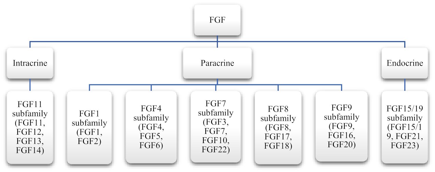 Fig.1 FGFs' classification according to their mode of actions. (Farooq M, et al., 2021)
FGFs bind to Fibroblast Growth Factor Receptors (FGFRs) and activate downstream signaling pathways. FGFs play essential roles in development, tissue repair, and cellular processes. Dysregulation of FGF signaling is associated with various diseases, and targeting FGFs and FGFRs holds therapeutic potential. Understanding the functions and mechanisms of FGF signaling contributes to our knowledge of normal physiology and disease pathogenesis.
Biological Functions and Mechanisms of Action of FGF
FGFs are involved in a wide range of biological functions, and their mechanisms of action are diverse and intricate. Here are some key biological functions and mechanisms of FGF:
- Cellular Proliferation and Survival: FGFs promote cell division and proliferation in various cell types. They stimulate the cell cycle progression by activating signaling pathways that drive cell growth and DNA replication. FGFs also play a role in cell survival by preventing apoptosis (programmed cell death) in certain cell populations.
- Angiogenesis: FGFs are potent angiogenic factors, meaning they stimulate the formation of new blood vessels. They promote endothelial cell proliferation and migration, which are essential steps in the process of angiogenesis. FGFs also induce the production of other angiogenic factors and extracellular matrix components necessary for blood vessel development.
- Tissue Development and Differentiation: FGFs participate in various stages of organogenesis, including the formation and patterning of tissues and organs. FGFs control cell fate determination and differentiation of specific cell lineages during development.
- Wound Healing and Tissue Repair: FGFs stimulate the proliferation of fibroblasts and other cell types involved in tissue repair, such as endothelial cells and keratinocytes. FGFs also promote the production of extracellular matrix components, facilitating tissue remodeling and scar formation.
- Neurogenesis and Neural Functions: FGFs regulate neurogenesis (generation of new neurons), axonal growth, and neuronal survival. FGFs also play a role in synaptic plasticity and neurotransmitter release, impacting neuronal communication and synaptic functions.
- Metabolism and Hormonal Regulation: FGFs, particularly FGF19, FGF21, and FGF23, have been implicated in metabolic regulation and hormone signaling. They influence glucose and lipid metabolism, energy homeostasis, and bile acid synthesis. FGF21, in particular, has been associated with the regulation of insulin sensitivity, adipogenesis, and thermogenesis.
The mechanisms of action of FGFs involve binding to specific cell surface receptors (FGFRs).
- Upon FGF binding, FGFRs undergo dimerization or oligomerization, which leads to the activation of their intracellular tyrosine kinase domains. This activation results in the autophosphorylation of tyrosine residues within the receptors.
- Phosphorylated tyrosine residues serve as docking sites for various intracellular signaling molecules, including adaptor proteins and kinases. These molecules initiate downstream signaling pathways, such as the MAPK pathway, PI3K/Akt pathway, and STAT pathway. These pathways regulate gene expression, protein synthesis, and other cellular processes, ultimately mediating the biological effects of FGFs.
- In addition to signaling through FGFRs, FGFs can interact with heparan sulfate proteoglycans (HSPGs), which act as co-receptors. HSPGs facilitate the binding of FGFs to FGFRs and stabilize the FGF-FGFR complex, enhancing the signaling response.
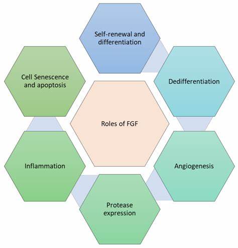 Fig.2 Role of FGF in tissue repair processes. (Farooq M, et al., 2021)
Understanding the diverse functions and mechanisms of FGF signaling is crucial for deciphering their roles in normal physiology and disease pathogenesis and for developing therapeutic interventions targeting FGF signaling pathways.
If you have any questions, requirements, or cooperation intentions, please feel free to contact us. We very much look forward to working with you and helping you achieve research and commercial success.
Related References
- Farooq M, Khan AW, Kim MS, Choi S. The Role of Fibroblast Growth Factor (FGF) Signaling in Tissue Repair and Regeneration. Cells. 2021; 10(11):3242.
- Deng J, Liu Y, Liu Y, Li W, Nie X. The Multiple Roles of Fibroblast Growth Factor in Diabetic Nephropathy. J Inflamm Res. 2021;14:5273-5290.