


gamma delta T Cells
Related Symbol Search List
- CD40
- CXCL13
- CXCR4
- ICOS
- CD27
- CD40 Ligand
- CD70
- CD4
- KLRC3
- FASLG
- GZMA
- Granzyme B
- Ifng
- TNFa
- CCL2
- CCR5
- CCR6
- CD3e
- CD83
- CD8A
- E-Cadherin
- Dectin-1
- Csf2
- DEFA1
- DEFB103B
- DEFB4A
- EGF
- EOMES
- Fas
- CD16a
- FCGR3B
- FGF10
- FGF7
- GATA3
- GNLY
- IGF1
- IL-10
- IL12A
- IL12B
- IL-13
- IL17A
- IL18R1
- IL2
- IL22
- IL23R
- IL-4
- Il5
- Il6
- KLRB1
- NKG2D
- OCLN
- RORC
- TARP
- TBX21
- TLR2
- TNFSF10
- ZBTB16
Immunology Background
Available Resources Related to Gamma Delta T Cells
Creative BioMart offers a wide variety of products, customized services, and resources dedicated to γδ T cell research. Our goal is to provide you with the tools and support necessary to advance your understanding of γδ T cell molecules and their role in the immune response.
We offer a wide range of high-quality products designed to support γδ T cell research. Our portfolio includes antibodies, recombinant proteins, protein-coupled magnetic beads, cell and tissue lysates, chromatography reagents, assay kits, and others. |
We understand that every research project is unique. We offer a range of services customized to your specific γδ T cell research needs. |
We offer a wide range of resources related to γδ T cell molecules, including protein functions, protein interactions, involved pathways, and other valuable information, providing researchers with strong support to help advance their research programs and experiments. |
About Gamma Delta T Cells
Gamma delta T cells, also known as γδ T cells, are a subset of T lymphocytes that possess a unique T cell receptor (TCR) composed of gamma (γ) and delta (δ) chains. Unlike the more common alpha-beta (αβ) T cells, which constitute the majority of T cells in the immune system, γδ T cells are a relatively small population, accounting for only 1-5% of circulating T cells in humans.
γδ T cells can be classified into subpopulations based on T cell receptor (TCR) usage, tissue distribution, and functional characteristics. the classification of γδ T cells may vary between species. The following is the most recent classification of γδ T cells taking into account the human and mouse γδ T cell subpopulations:
| Human γδ T cell subsets |
|
| Mouse γδ T cell subsets |
|
One of the defining features of γδ T cells is their ability to recognize a diverse range of antigens, including both protein and non-protein antigens. While αβ T cells primarily recognize peptide antigens presented by major histocompatibility complex (MHC) molecules, γδ T cells can directly recognize antigens without requiring MHC presentation. This enables them to respond rapidly to stress-induced molecules, such as certain microbial components, tumor-associated antigens, and damaged or infected cells.
γδ T cells exhibit a wide range of effector functions and can contribute to both innate and adaptive immune responses. They can produce various cytokines, including interleukin-17 (IL-17), interferon-gamma (IFN-γ), and tumor necrosis factor-alpha (TNF-α), which are involved in inflammatory responses, antimicrobial defense, and tissue repair. Additionally, γδ T cells can directly kill infected or malignant cells through cytotoxic mechanisms.
Furthermore, γδ T cells have been implicated in immune regulation and modulation of other immune cells. They can interact with dendritic cells, B cells, and other T cell subsets, influencing the overall immune response and contributing to the balance between tolerance and immunity.
Due to their unique properties and functions, γδ T cells have gained increasing attention in the field of immunology and their potential therapeutic applications are being explored. Understanding the biology of γδ T cells and their interactions with other immune cells may provide valuable insights for the development of novel immunotherapies and vaccines targeting various diseases, including infections, cancer, and autoimmune disorders.
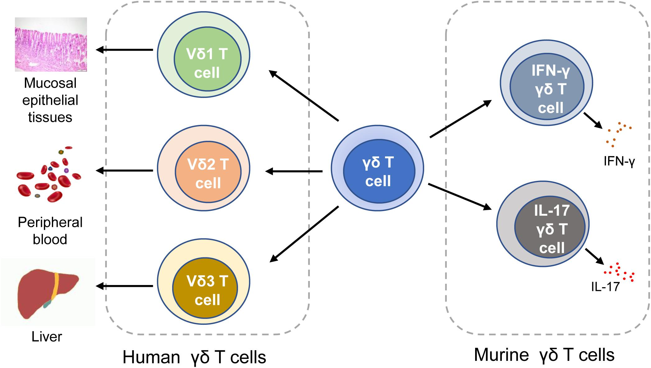 Fig.1 Classification of γδ T cells. In human, γδ T cells can be classified into Vδ1 T cells, Vδ1 T cells, and Vδ3 T cells. In mice, γδ T cells can be categorized into IFN-γγδ T cells and IL-17 γδ T cells. IFN-γ, interferon-γ. (Ma R, et al., 2020)
Mechanisms of γδ T Cell Activation and Regulation
| Key mechanisms name | Mechanisms |
|---|---|
| 1. TCR recognition of antigens | The activation of γδ T cells begins with the recognition of antigens by their T cell receptor (TCR). The γδ TCR is capable of recognizing a diverse range of antigens, including peptides, lipids, and phosphorylated metabolites, in a major histocompatibility complex (MHC)-independent manner. This antigen recognition triggers intracellular signaling cascades, leading to γδ T cell activation. |
| 2. Co-stimulation | Co-stimulatory signals are crucial for the full activation of γδ T cells. Co-stimulatory molecules, such as CD28 and CD27, interact with their ligands on antigen-presenting cells (APCs) or target cells, providing additional signals that enhance γδ T cell activation and effector functions. |
| 3. Cytokines | Various cytokines, including interleukin-2 (IL-2), interleukin-7 (IL-7), and interleukin-15 (IL-15), are required for the survival, proliferation, and differentiation of γδ T cells. These cytokines are produced by APCs, stromal cells, or other immune cells in the microenvironment. |
| 4. Antigen-presenting cells (APCs) | Professional APCs, such as dendritic cells, macrophages, and B cells, can present antigens to γδ T cells. Upon antigen recognition, APCs provide co-stimulatory signals and secrete cytokines that influence the activation and differentiation of γδ T cells. |
| 5. Recognition of stress-induced molecules | γδ T cells can detect stress-induced molecules or danger-associated molecular patterns (DAMPs) released by infected or damaged cells. These molecules, such as heat shock proteins or phosphoantigens, can be recognized by specific receptors on γδ T cells, such as Toll-like receptors (TLRs) or γδ TCRs, leading to activation. |
| 6. Regulation by inhibitory receptors | γδ T cells express inhibitory receptors that regulate their activation and effector functions. For example, immune checkpoint molecules like programmed cell death protein 1 (PD-1) or cytotoxic T-lymphocyte-associated protein 4 (CTLA-4) can limit the activation and suppressive capacity of γδ T cells. |
| 7. Regulatory T cell interactions | Regulatory T cells (Tregs) can interact with γδ T cells and influence their activation and functions. Tregs can inhibit the activation of γδ T cells through cytokine-mediated or cell contact-dependent mechanisms, contributing to immune regulation and tolerance. |
| 8. Tissue microenvironment | The specific tissue microenvironment and local cues influence γδ T cell activation and regulation. Epithelial tissues, such as skin or mucosal surfaces, provide unique signals that shape the phenotype and function of resident γδ T cells. |
| 9. Epigenetic regulation | Epigenetic mechanisms, such as DNA methylation or histone modifications, can regulate the activation and differentiation of γδ T cells. Epigenetic changes can influence the expression of genes involved in γδ T cell activation and effector functions. |
Role of γδ T Cell-Associated Molecules in Immune Regulation and Immune Response
The molecules associated with γδ T cells play crucial roles in immune regulation and immune responses. Here are some key molecules and their functions:
- T cell receptor (TCR): The γδ TCR is responsible for antigen recognition by γδ T cells. It binds to specific antigens, initiating signaling cascades that lead to γδ T cell activation and effector functions. The diversity of γδ TCR repertoire allows for the recognition of a wide range of antigens, including both protein and non-protein antigens.
- Co-stimulatory molecules: Co-stimulatory molecules provide additional signals required for full activation of γδ T cells. Examples include CD28 and CD27, which can interact with their respective ligands on antigen-presenting cells or target cells. Co-stimulation enhances γδ T cell responses and promotes their effector functions.
- Cytokines: Cytokines are key regulators of immune responses, and γδ T cells can produce various cytokines with immunoregulatory functions. For instance, γδ T cells can produce interferon-gamma (IFN-γ), which promotes inflammation, enhances antimicrobial activity, and modulates immune cell functions. They can also produce interleukin-17 (IL-17), which is involved in pro-inflammatory responses and the recruitment of neutrophils.
- Natural Killer (NK) receptors: Some γδ T cells express NK receptors, such as NKG2D. These receptors can recognize stress-induced ligands or molecules expressed on infected or transformed cells. Engagement of NK receptors on γδ T cells can trigger cytotoxicity and the release of cytotoxic molecules, contributing to the elimination of target cells.
- Adhesion molecules: Adhesion molecules facilitate the interaction between γδ T cells and other immune cells or target cells. For example, integrins such as LFA-1 (lymphocyte function-associated antigen-1) and CD2 are important for cell-cell adhesion and migration of γδ T cells to inflamed tissues or lymphoid organs.
- Cytotoxic molecules: γδ T cells can directly kill target cells through the release of cytotoxic molecules. These include perforin, which forms pores in the target cell membrane, and granzymes, which induce apoptosis in the target cells. The expression of cytotoxic molecules allows γδ T cells to eliminate infected cells or tumor cells.
- Regulatory molecules: γδ T cells can express various regulatory molecules that modulate immune responses. For example, they may express immune checkpoint molecules, such as PD-1 (programmed cell death protein 1) or CTLA-4 (cytotoxic T-lymphocyte-associated protein 4), which regulate the activity of γδ T cells and prevent excessive immune activation.
These molecules collectively contribute to the immunoregulatory and effector functions of γδ T cells. They enable γδ T cells to recognize specific antigens, activate downstream signaling pathways, interact with other immune cells, produce cytokines, exert cytotoxicity, and participate in immune regulation. The precise role of these molecules can vary depending on the specific context, the antigen encountered, and the overall immune response.
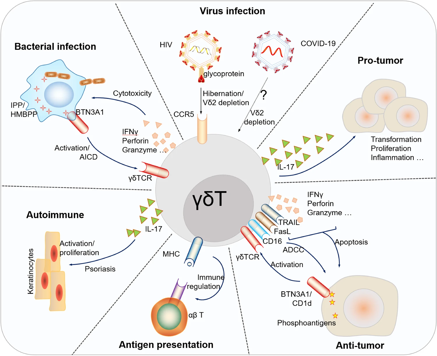 Fig.2 Brief sketch depicts the major roles of human γδ T cells in the immune regulation, pathogenesis and progression of diverse diseases (representative mechanisms shown). (Hu, Y., et al., 2023)
Role of γδ T-Cell-Associated Molecules in Disease Onset and Progression
The molecules associated with γδ T cells can play important roles in the onset and progression of various diseases. Here are some examples of how γδ T-cell-associated molecules contribute to disease:
- Autoimmune diseases: In autoimmune diseases, γδ T-cell-associated molecules can contribute to the dysregulation of immune responses leading to tissue damage. For instance, γδ T cells expressing certain TCRs and cytokines, such as IL-17, have been implicated in the pathogenesis of autoimmune diseases like psoriasis, rheumatoid arthritis, and inflammatory bowel disease. These γδ T cells can promote chronic inflammation and induce tissue destruction.
- Infectious diseases: γδ T cells and their associated molecules play a crucial role in the immune response against various pathogens. They can recognize and respond to specific antigens derived from bacteria, viruses, and parasites. The activation of γδ T cells through their TCRs and the production of cytokines, such as IFN-γ, can contribute to the clearance of infections. However, dysregulated or excessive γδ T cell responses can also contribute to immunopathology in certain infectious diseases, such as tuberculosis or malaria.
- Cancer: γδ T cells and their associated molecules have gained attention as potential mediators of anti-tumor immune responses. γδ T cells expressing specific TCRs can recognize tumor-associated antigens and directly kill cancer cells. The expression of cytotoxic molecules, such as perforin and granzymes, by activated γδ T cells contributes to their anti-tumor cytotoxicity. Additionally, γδ T cells can produce cytokines, such as IFN-γ and TNF-α, which can exert anti-tumor effects by inhibiting tumor growth and promoting immune cell activation.
- Allergic diseases: In allergic diseases, γδ T cells and their associated molecules can contribute to the immune response and tissue inflammation. For example, γδ T cells expressing certain TCRs, such as Vγ4Vδ5, have been implicated in allergic contact dermatitis. These cells can recognize specific allergens and release cytokines, such as IL-17 and IL-22, which promote inflammation and tissue damage.
- Transplant rejection: γδ T cells and their associated molecules can contribute to the rejection of transplanted organs. γδ T cells can recognize and respond to non-self antigens presented by MHC molecules on transplanted tissues. The activation of γδ T cells can lead to the production of pro-inflammatory cytokines, recruitment of other immune cells, and tissue damage, ultimately leading to graft rejection.
- Neurological disorders: Emerging evidence suggests that γδ T cells and their associated molecules may play a role in the pathogenesis of certain neurological disorders, such as multiple sclerosis and Alzheimer's disease. γδ T cells can infiltrate the central nervous system and contribute to neuroinflammation through the production of pro-inflammatory cytokines and interaction with other immune cells.
It is important to note that the specific roles of γδ T-cell-associated molecules in disease onset and progression can vary depending on the disease context, the specific γδ T-cell subset involved, and the overall immune response. The dysregulation or imbalance in the activation or function of γδ T cells and their associated molecules can contribute to disease pathogenesis, highlighting their potential as targets for therapeutic interventions.
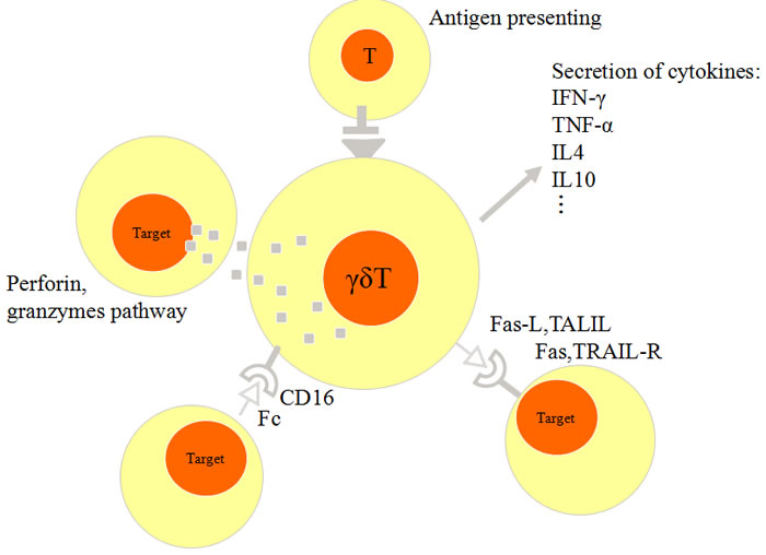 Fig.3 Schematic figure of anti-tumor activity of γδ T cells. 1) γδ T cells secret IFN-γ and TNF-α, IL-4 and IL-10. 2) enhanced expression of Fas-L and TRAIL in γδ T cells. 3) γδ T cells express CD16 mediates Fc antibody-dependent cellular cytotoxicity (ADCC). 4) γδ T cells release perforin and granzymes for cytotoxic activity. (Zou C, et al., 2017)
For more information, please contact our team of experts for personalized assistance. Explore our products today to unlock the full potential of γδ T cells in immune response and disease understanding! Let's drive innovation together and achieve breakthroughs in γδ T cell research!
Related References
- Ma R, Yuan D, Guo Y, Yan R, Li K. Immune Effects of γδ T Cells in Colorectal Cancer: A Review. Front Immunol. 2020;11:1600.
- Hu, Y., Hu, Q., Li, Y. et al. γδ T cells: origin and fate, subsets, diseases and immunotherapy. Sig Transduct Target Ther 8, 434 (2023).
- Zou C, Zhao P, Xiao Z, Han X, Fu F, Fu L. γδ T cells in cancer immunotherapy. Oncotarget. 2017;8(5):8900-8909.