


T Follicular Helper (Tfh) Cells
Related Symbol Search List
- CR2
- CR1
- SLAMF7
- VAV1
- BTLA
- CD28
- CD40
- CD80
- CD84
- CD86
- Icosl
- LY9
- PDCD1
- Tnfsf4
- CD32A
- CCL19
- CCL21
- CXCL13
- CXCR4
- CXCR5
- BATF
- PRDM1
- CD274
- ICOS
- PD-L2
- CD40 Ligand
- TNFRSF4
- TNFRSF8
- TNFSF8
- CD4
- CCR7
- Ifng
- Fas
- GATA3
- IL-10
- IL-13
- IL2
- IL-4
- Il5
- Il6
- KLRB1
- BCL6
- CD109
- CD200
- CD3G
- CD69
- IFNGR1
- IL10RA
- IL10RB
- IL17RA
- IL21
- IL21R
- IL2RA
- IL2RB
- IL4R
- IL5RA
- Il6ra
- IL6ST
- INPP5D
- INPPL1
- JAK1
- CD10
- NFATC1
- PIK3CB
- PIK3CD
- PIK3R1
- PIK3R2
- PTEN
- STAT3
- TYK2
Immunology Background
Available Resources Related to T Follicular Helper (Tfh) Cells
Creative BioMart is dedicated to providing a broad range of products, custom services, and resources to support researchers studying T follicular helper (Tfh) cells. Our goal is to advance the understanding of Tfh cell biology and their role in various immune processes.
| We offer a comprehensive range of high-quality products designed specifically for Tfh cell research. Our product portfolio includes recombinant proteins, GMP proteins, protein-coupled magnetic beads, cell and tissue lysates, chromatography reagents, antibodies, assay kits, and others. | |
| We understand that every research project has unique requirements. Therefore, we offer services tailored to your specific needs, including custom protein expression, custom antibody development, custom assay development, and more, to study Tfh cell biology and related immune responses. | |
| We are also committed to providing valuable resources and support to the scientific community, including protein functions, protein interactions, involved pathways, and other valuable information. Helping researchers gain a deeper understanding of Tfh cell function and its impact on a variety of immune-mediated diseases. |
About T Follicular Helper (Tfh) Cells
T follicular helper (Tfh) cells are a specialized subset of CD4+ T cells that play a crucial role in the generation and maintenance of high-affinity antibody responses. They are primarily found within the germinal centers of secondary lymphoid organs, such as lymph nodes and spleen, where they interact with B cells to support the production of long-lived antibody-secreting cells and memory B cells. Tfh cells provide critical help to B cells in the process of affinity maturation and class switching, ultimately leading to the generation of protective antibodies.
Characteristics and Phenotype
Tfh cells can be identified by the expression of specific cell surface markers and transcription factors. They typically express the chemokine receptor CXCR5, which guides their migration toward the B cell follicles, where they form stable interactions with B cells. Tfh cells also display high levels of the co-stimulatory molecule ICOS (inducible T cell co-stimulator) and the surface protein PD-1 (programmed cell death protein 1).
Transcriptionally, Tfh cells are defined by the expression of the master transcription factor Bcl-6 (B-cell lymphoma 6), which is critical for their development and function. Bcl-6 regulates the expression of various molecules involved in Tfh cell differentiation, migration, and interaction with B cells.
Role in B Cell Help
Tfh cells provide essential help to B cells at multiple stages of the immune response:
- Germinal Center Formation: Tfh cells migrate to the B cell follicles in secondary lymphoid organs and promote the formation of germinal centers, specialized structures where B cells undergo somatic hypermutation and affinity maturation.
- B Cell Activation and Differentiation: Tfh cells interact with B cells through cell surface molecules, such as CD40 ligand (CD40L) and ICOS ligand, and provide signals for B cell activation, survival, and differentiation. These interactions stimulate B cells to undergo class switching to produce different antibody isotypes (e.g., IgG, IgA) and promote the generation of plasma cells and memory B cells.
- Selection of High-Affinity B Cells: Tfh cells contribute to the selection of B cells with high-affinity B cell receptors (BCRs) through a process known as "affinity maturation." Tfh cells provide signals that promote the survival and proliferation of B cells with higher affinity BCRs, leading to the production of antibodies with increased antigen specificity.
Regulation and Dysregulation
The differentiation and function of Tfh cells are tightly regulated to ensure an appropriate immune response. Several factors, including cytokines (such as IL-6, IL-21, and IL-12), transcription factors (e.g., Bcl-6, STAT3), and signaling pathways (e.g., PI3K-Akt-mTOR), influence Tfh cell development and maintenance.
Dysregulation of Tfh cells has been implicated in various immune-related disorders. Excessive Tfh cell activation and dysregulated germinal center responses have been associated with autoimmunity, such as systemic lupus erythematosus (SLE) and rheumatoid arthritis (RA). Conversely, deficiencies in Tfh cell function can lead to impaired antibody responses and increased susceptibility to infections.
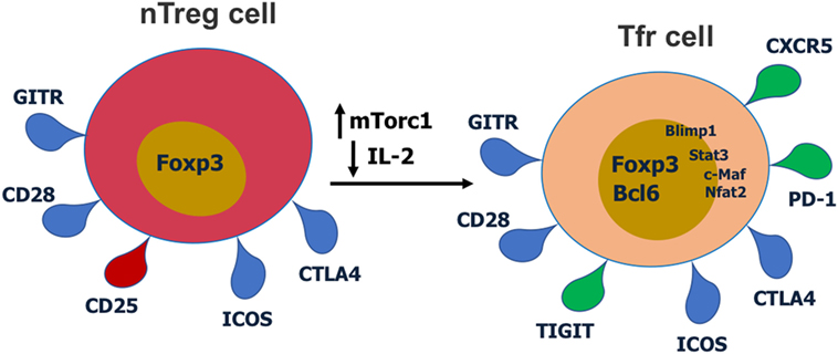 Fig.1 Cell surface receptors and transcription factors involved in T follicular regulatory (Tfr) cell differentiation and function. (Xie M M, et al., 2018)
Key Molecules Involved in the Function of Tfh Cells
Here is a list of key molecules involved in the function of Tfh cells and their roles in Tfh cell differentiation and function (These are just a few examples of the molecules associated with Tfh cells):
| Key molecules | Functions |
|---|---|
| CXCR5 (C-X-C chemokine receptor type 5) | CXCR5 is a chemokine receptor expressed on the surface of Tfh cells. It guides their migration towards the B cell follicles within secondary lymphoid organs, where they interact with B cells. CXCR5 helps Tfh cells localize to the germinal centers, facilitating their role in supporting B cell responses. |
| ICOS (Inducible T cell co-stimulator) | ICOS is a co-stimulatory molecule expressed on Tfh cells. Interaction between ICOS on Tfh cells and its ligand, ICOSL, on B cells provides crucial co-stimulatory signals necessary for Tfh cell differentiation and function. ICOS signaling promotes Tfh cell development, survival, and cytokine production. |
| PD-1 (Programmed cell death protein 1) | PD-1 is an immune checkpoint receptor expressed on Tfh cells. Engagement of PD-1 with its ligands, PD-L1 and PD-L2, regulates Tfh-B cell interactions within the germinal centers. PD-1 signaling helps maintain the balance between Tfh cell activation and immune tolerance, preventing excessive activation and autoimmune responses. |
| Bcl-6 (B-cell lymphoma 6) | Bcl-6 is a master transcription factor that plays a central role in Tfh cell differentiation and function. It is highly expressed in Tfh cells and regulates the expression of genes associated with Tfh cell development, migration, and effector functions. Bcl-6 promotes Tfh cell differentiation by suppressing alternative T helper cell lineages and orchestrates the expression of molecules involved in Tfh cell-B cell interactions. |
| IL-21 (Interleukin-21) | IL-21 is a cytokine produced by Tfh cells and is crucial for their differentiation and function. It acts in an autocrine manner to drive Tfh cell development and supports B cell responses within the germinal centers. IL-21 promotes B cell proliferation, class switching, and affinity maturation, ultimately leading to the production of high-affinity antibodies. |
| CD40L (CD40 ligand) | CD40L is a surface molecule expressed on Tfh cells that interacts with CD40 on B cells. This interaction provides essential co-stimulatory signals for B cell activation, differentiation, and antibody production. CD40L-CD40 signaling is critical for the formation and maintenance of germinal centers and supports Tfh cell-mediated help to B cells. |
| IL-6 (Interleukin-6) and IL-12 (Interleukin-12) | These cytokines, along with other signals, influence the differentiation of Tfh cells. IL-6 and IL-12 are involved in promoting Tfh cell differentiation by activating STAT3 and inducing Bcl-6 expression. They contribute to the development of Tfh cell effector functions and the generation of productive Tfh-B cell interactions. |
These molecules collectively contribute to Tfh cell differentiation, migration, and their interactions with B cells, facilitating the generation of robust antibody responses and germinal center reactions. Their precise regulation and coordination are essential for effective humoral immune responses and maintaining immune homeostasis.
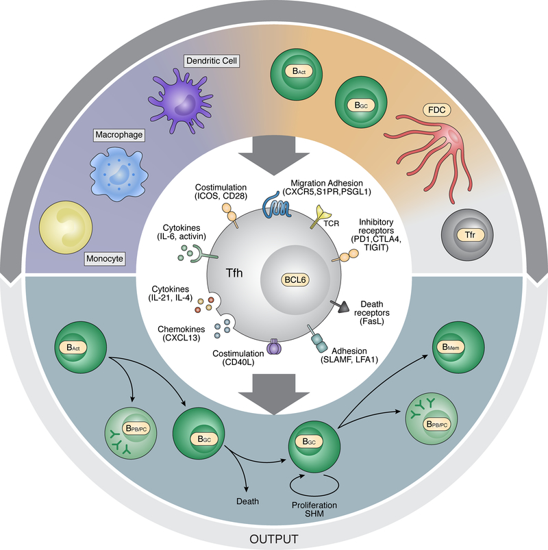 Fig.2 Signals to and from Tfh cells. Tfh cells receive signals from a number of cell types that can regulate Tfh differentiation and/or function. Tfh cells function by providing help to B cells at a series of stages of antigen-specific B cell differentiation. (Crotty S, 2019)
How Dysregulation of Tfh Cell-Related Molecules Can Contribute to Diseases?
Dysregulation of Tfh cell-related molecules can have significant implications for the development and progression of autoimmune diseases, allergies, and other immune-related disorders. Here's some information on how dysregulation of these molecules can contribute to these conditions:
Autoimmune Diseases
Autoimmune diseases occur when the immune system mistakenly attacks the body's own tissues. Dysregulation of Tfh cell-related molecules can contribute to the development of autoimmune diseases in the following ways:
- Reduced PD-1 expression or impaired PD-1 signaling can lead to unchecked Tfh cell activation and excessive germinal center reactions. This dysregulation can contribute to the breakdown of immune tolerance and the production of pathogenic autoantibodies, as seen in systemic lupus erythematosus (SLE) and rheumatoid arthritis (RA).
- Aberrant expression or dysregulation of Bcl-6 can disrupt the balance between Tfh and other T cell lineages, leading to the accumulation of autoreactive Tfh cells. These autoreactive Tfh cells can drive the production of autoantibodies and contribute to autoimmune disease development.
Allergies and Allergic Diseases
Allergies and allergic diseases occur when the immune system overreacts to harmless substances. Dysregulation of Tfh cell-related molecules can influence the development and severity of allergic responses:
- Excessive ICOS signaling and IL-21 production by Tfh cells can contribute to the generation of excessive B cell responses and the production of allergen-specific antibodies in allergic diseases such as allergic asthma and allergic rhinitis.
- Dysregulated CXCR5 expression or signaling can affect the migration and localization of Tfh cells within lymphoid tissues. This alteration may disrupt the interaction between Tfh cells and B cells, impairing the generation of appropriate immune responses against allergens.
Other Immune-Related Disorders
Dysregulation of Tfh cell-related molecules can also contribute to other immune-related disorders beyond autoimmune diseases and allergies:
- Imbalanced production or dysregulated signaling of IL-6 and IL-12 can affect Tfh cell differentiation and function. Altered levels of these cytokines have been implicated in immune-related disorders such as graft-versus-host disease (GVHD) and certain types of immunodeficiency.
- Defects in CD40L expression or signaling can lead to impaired Tfh cell-mediated help to B cells, resulting in compromised antibody responses and increased susceptibility to infections.
It is important to note that dysregulation of Tfh cell-related molecules often occurs in conjunction with other genetic, environmental, and immunological factors, contributing to the complex pathogenesis of these immune-related disorders. Further research is needed to fully understand the mechanisms underlying the dysregulation of Tfh cell-related molecules and their precise roles in the development and progression of these conditions.
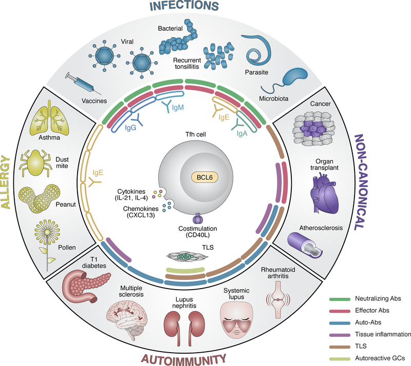 Fig.3 Relationships of Tfh cells to diseases. (Crotty S, 2019)
Our team of scientists is always available to provide you with expert guidance and assistance. Whether you need help with experimental design, product selection, or troubleshooting, we are here to support your Tfh cell research endeavors. For more information, please contact our team of experts for personalized assistance. Let's drive innovation together and achieve breakthroughs in Tfh cell research!
Related References
- Xie M M, Dent A L. Unexpected help: follicular regulatory T cells in the germinal center[J]. Frontiers in immunology, 2018, 9: 387906.
- Crotty S. T Follicular Helper Cell Biology: A Decade of Discovery and Diseases. Immunity. 2019;50(5):1132-1148.
- Gutiérrez-Melo N, Baumjohann D. T follicular helper cells in cancer. Trends Cancer. 2023;9(4):309-325.
- Ma CS, Deenick EK. Human T follicular helper (Tfh) cells and disease. Immunol Cell Biol. 2014;92(1):64-71.